Stockmeier Craig A, Mahajan Gouri J, Konick Lisa C, Overholser James C, Jurjus George J, Meltzer Herbert Y, Uylings Harry B M, Friedman Lee, Rajkowska Grazyna
Department of Psychiatry and Human Behavior (Box 127), University of Mississippi Medical Center, 2500 North State Street, Jackson, MS 39216, USA.
Biol Psychiatry. 2004 Nov 1;56(9):640-50. doi: 10.1016/j.biopsych.2004.08.022.
Imaging studies report that hippocampal volume is decreased in major depressive disorder (MDD). A cellular basis for reduced hippocampal volume in MDD has not been identified.
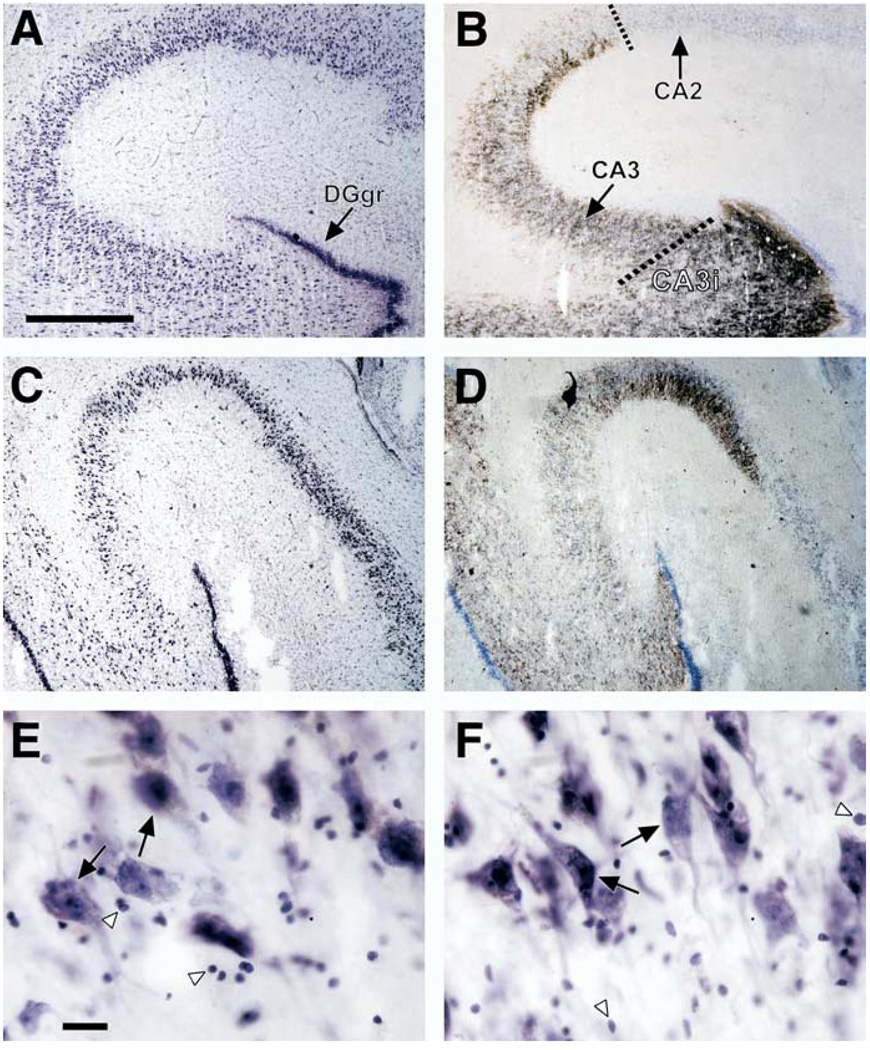
Sections of right hippocampus were collected in 19 subjects with MDD and 21 normal control subjects. The density of pyramidal neurons, dentate granule cell neurons, glia, and the size of the neuronal somal area were measured in systematic, randomly placed three-dimensional optical disector counting boxes.
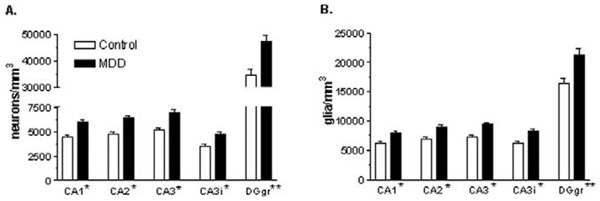
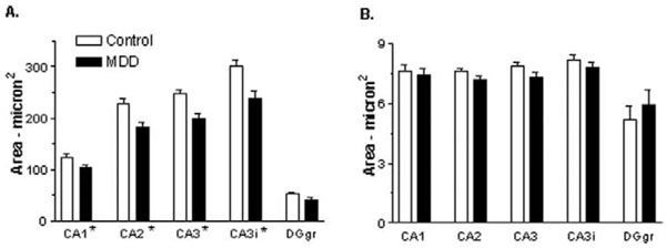
In MDD, cryostat-cut hippocampal sections shrink in depth a significant 18% greater amount than in control subjects. The density of granule cells and glia in the dentate gyrus and pyramidal neurons and glia in all cornv ammonis (CA)/hippocampal subfields is significantly increased by 30%-35% in MDD. The average soma size of pyramidal neurons is significantly decreased in MDD.
In MDD, the packing density of glia, pyramidal neurons, and granule cell neurons is significantly increased in all hippocampal subfields and the dentate gyrus, and pyramidal neuron soma size is significantly decreased as well. It is suggested that a significant reduction in neuropil in MDD may account for decreased hippocampal volume detected by neuroimaging. In addition, differential shrinkage of frozen sections of the hippocampus suggests differential water content in hippocampus in MDD.
影像学研究报告称,重度抑郁症(MDD)患者的海马体体积减小。MDD中海马体体积减小的细胞基础尚未明确。
收集了19名MDD患者和21名正常对照者的右侧海马体切片。在系统随机放置的三维光学分割计数框中测量锥体细胞、齿状颗粒细胞神经元、神经胶质细胞的密度以及神经元胞体面积大小。
在MDD患者中,低温恒温器切割的海马体切片深度收缩比对照者显著大18%。MDD患者齿状回中的颗粒细胞和神经胶质细胞以及所有海马旁回(CA)/海马亚区中的锥体细胞和神经胶质细胞密度显著增加30%-35%。MDD患者锥体细胞的平均胞体大小显著减小。
在MDD中,所有海马亚区和齿状回中神经胶质细胞、锥体细胞和颗粒细胞神经元的堆积密度显著增加,锥体细胞胞体大小也显著减小。提示MDD中神经毡的显著减少可能是神经影像学检测到的海马体体积减小的原因。此外,海马体冷冻切片的差异收缩表明MDD患者海马体中的含水量存在差异。