• 文献检索
  • 文档翻译
  • 深度研究
  • 学术资讯
  • Suppr Zotero 插件Zotero 插件
  • 邀请有礼
  • 套餐&价格
  • 历史记录
应用&插件
Suppr Zotero 插件Zotero 插件浏览器插件Mac 客户端Windows 客户端微信小程序
定价
高级版会员购买积分包购买API积分包
服务
文献检索文档翻译深度研究API 文档MCP 服务
关于我们
关于 Suppr公司介绍联系我们用户协议隐私条款
关注我们

Suppr 超能文献

核心技术专利:CN118964589B侵权必究
粤ICP备2023148730 号-1Suppr @ 2026

文献检索

告别复杂PubMed语法,用中文像聊天一样搜索,搜遍4000万医学文献。AI智能推荐,让科研检索更轻松。

立即免费搜索

文件翻译

保留排版,准确专业,支持PDF/Word/PPT等文件格式,支持 12+语言互译。

免费翻译文档

深度研究

AI帮你快速写综述,25分钟生成高质量综述,智能提取关键信息,辅助科研写作。

立即免费体验

The use of Raman spectroscopy to differentiate between different prostatic adenocarcinoma cell lines.

作者信息

Crow P, Barrass B, Kendall C, Hart-Prieto M, Wright M, Persad R, Stone N

机构信息

Biophotonics Research Group, Pullman Court, Gloucestershire Royal Hospital, Great Western Road, Gloucester GL1 3NN, UK.

出版信息

Br J Cancer. 2005 Jun 20;92(12):2166-70. doi: 10.1038/sj.bjc.6602638.

DOI:10.1038/sj.bjc.6602638
PMID:15928665
原文链接:https://pmc.ncbi.nlm.nih.gov/articles/PMC2361812/
Abstract

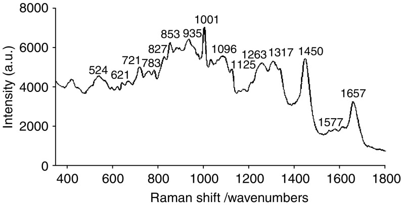
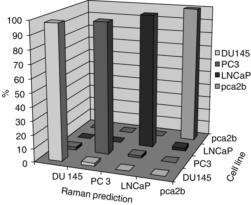
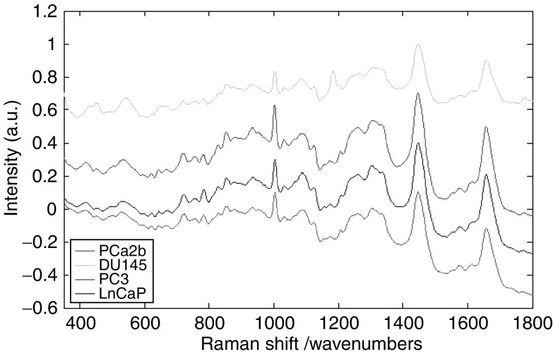
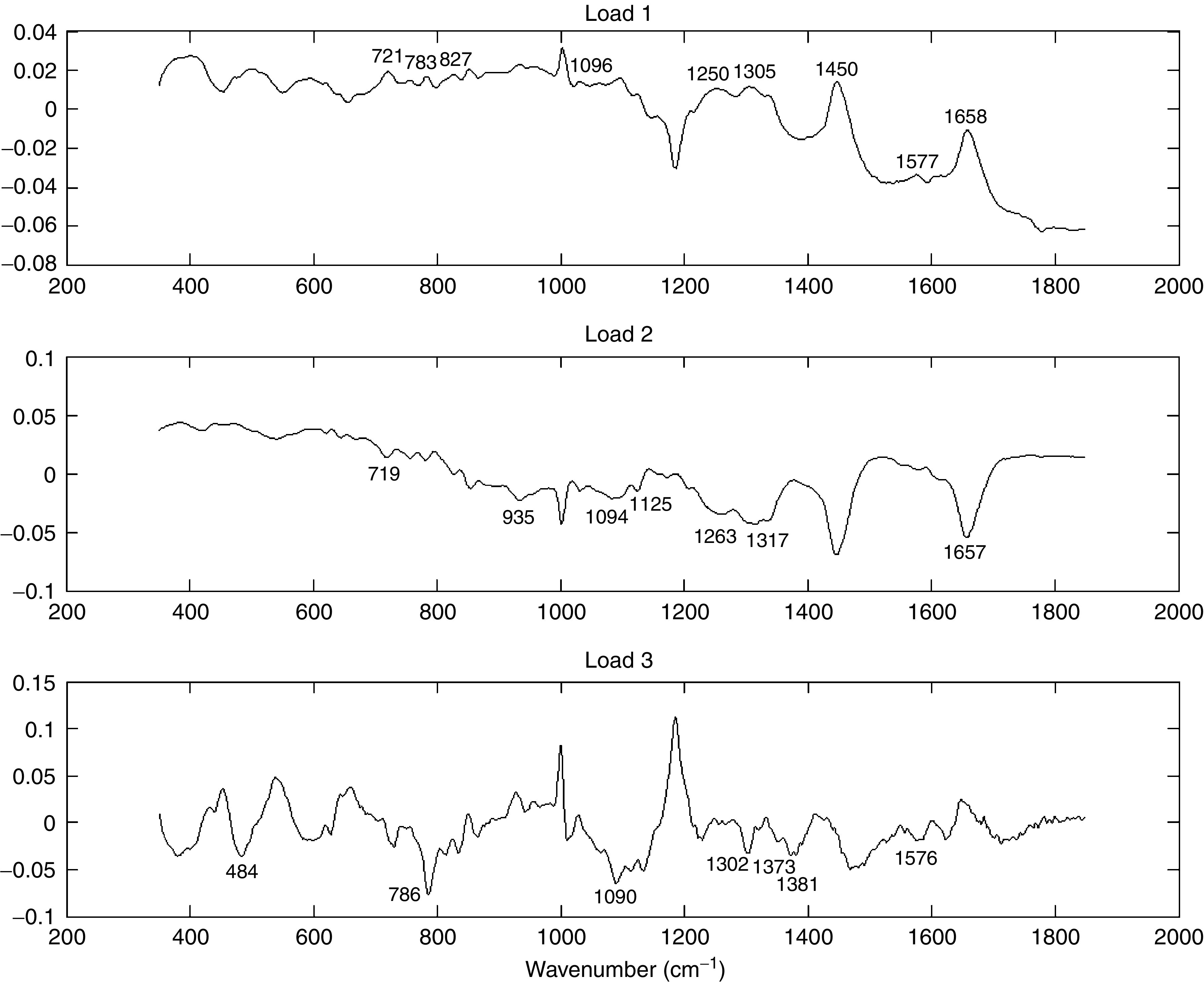
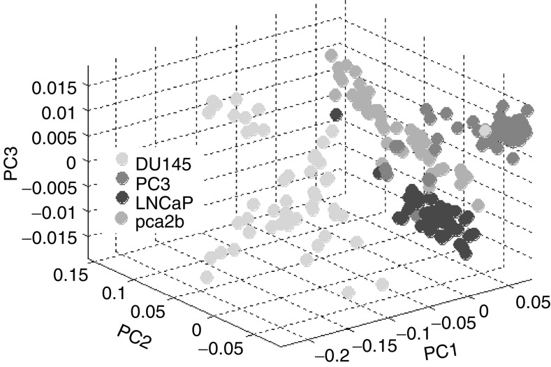
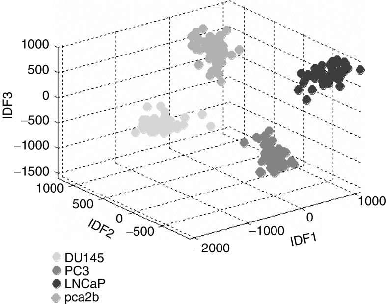
Raman spectroscopy (RS) is an optical technique that provides an objective method of pathological diagnosis based on the molecular composition of tissue. Studies have shown that the technique can accurately identify and grade prostatic adenocarcinoma (CaP) in vitro. This study aimed to determine whether RS was able to differentiate between CaP cell lines of varying degrees of biological aggressiveness. Raman spectra were measured from two well-differentiated, androgen-sensitive cell lines (LNCaP and PCa 2b) and two poorly differentiated, androgen-insensitive cell lines (DU145 and PC 3). Principal component analysis was used to study the molecular differences that exist between cell lines and, in conjunction with linear discriminant analysis, was applied to 200 spectra to construct a diagnostic algorithm capable of differentiating between the different cell lines. The algorithm was able to identify the cell line of each individual cell with an overall sensitivity of 98% and a specificity of 99%. The results further demonstrate the ability of RS to differentiate between CaP samples of varying biological aggressiveness. RS shows promise for application in the diagnosis and grading of CaP in clinical practise as well as providing molecular information on CaP samples in a research setting.

摘要
https://cdn.ncbi.nlm.nih.gov/pmc/blobs/1caa/2361812/466772e39b83/92-6602638f7.jpg
https://cdn.ncbi.nlm.nih.gov/pmc/blobs/1caa/2361812/9135fa9387c6/92-6602638f1.jpg
https://cdn.ncbi.nlm.nih.gov/pmc/blobs/1caa/2361812/975baed2cd4e/92-6602638f2.jpg
https://cdn.ncbi.nlm.nih.gov/pmc/blobs/1caa/2361812/3923a7db4404/92-6602638f3.jpg
https://cdn.ncbi.nlm.nih.gov/pmc/blobs/1caa/2361812/e8d9201a214b/92-6602638f4.jpg
https://cdn.ncbi.nlm.nih.gov/pmc/blobs/1caa/2361812/29c42f9059ee/92-6602638f5.jpg
https://cdn.ncbi.nlm.nih.gov/pmc/blobs/1caa/2361812/e7f8a8cd8861/92-6602638f6.jpg
https://cdn.ncbi.nlm.nih.gov/pmc/blobs/1caa/2361812/466772e39b83/92-6602638f7.jpg

相似文献

1
The use of Raman spectroscopy to differentiate between different prostatic adenocarcinoma cell lines.
Br J Cancer. 2005 Jun 20;92(12):2166-70. doi: 10.1038/sj.bjc.6602638.
2
The use of Raman spectroscopy to identify and grade prostatic adenocarcinoma in vitro.利用拉曼光谱在体外鉴定前列腺腺癌并进行分级。
Br J Cancer. 2003 Jul 7;89(1):106-8. doi: 10.1038/sj.bjc.6601059.
3
Biochemical signatures of in vitro radiation response in human lung, breast and prostate tumour cells observed with Raman spectroscopy.拉曼光谱观察到的人肺、乳腺和前列腺肿瘤细胞体外辐射反应的生化特征。
Phys Med Biol. 2011 Nov 7;56(21):6839-55. doi: 10.1088/0031-9155/56/21/006. Epub 2011 Oct 5.
4
Mesoscopic characterization of prostate cancer using Raman spectroscopy: potential for diagnostics and therapeutics.利用拉曼光谱对前列腺癌进行介观特征分析:在诊断和治疗方面的潜力。
BJU Int. 2018 Aug;122(2):326-336. doi: 10.1111/bju.14199. Epub 2018 Apr 13.
5
Assessment of fiberoptic near-infrared raman spectroscopy for diagnosis of bladder and prostate cancer.用于诊断膀胱癌和前列腺癌的光纤近红外拉曼光谱评估。
Urology. 2005 Jun;65(6):1126-30. doi: 10.1016/j.urology.2004.12.058.
6
Classification of fixed urological cells using Raman tweezers.使用拉曼镊子对固定泌尿细胞进行分类。
J Biophotonics. 2009 Feb;2(1-2):47-69. doi: 10.1002/jbio.200810061.
7
Raman spectroscopy, a potential tool in diagnosis and prognosis of castration-resistant prostate cancer.拉曼光谱学,一种在去势抵抗性前列腺癌的诊断和预后中有应用潜力的工具。
J Biomed Opt. 2013 Aug;18(8):87001. doi: 10.1117/1.JBO.18.8.087001.
8
Raman microscopy for the chemometric analysis of tumor cells.用于肿瘤细胞化学计量分析的拉曼显微镜技术。
J Phys Chem B. 2006 Oct 5;110(39):19625-31. doi: 10.1021/jp061981q.
9
The use of Raman spectroscopy to identify and characterize transitional cell carcinoma in vitro.利用拉曼光谱在体外鉴定和表征移行细胞癌。
BJU Int. 2004 Jun;93(9):1232-6. doi: 10.1111/j.1464-410X.2004.04852.x.
10
Diagnosis of cervical squamous cell carcinoma and cervical adenocarcinoma based on Raman spectroscopy and support vector machine.基于拉曼光谱和支持向量机诊断宫颈鳞状细胞癌和宫颈腺癌。
Photodiagnosis Photodyn Ther. 2019 Sep;27:156-161. doi: 10.1016/j.pdpdt.2019.05.029. Epub 2019 May 25.

引用本文的文献

1
Raman micro-spectroscopy reveals the metabolic alterations in primary prostate tumor tissues of patients with metastases.拉曼显微光谱揭示了有转移的前列腺癌患者原发性肿瘤组织中的代谢改变。
J Transl Med. 2025 Jun 17;23(1):675. doi: 10.1186/s12967-025-06655-4.
2
Unsupervised self-organising map classification of Raman spectra from prostate cell lines uncovers substratified prostate cancer disease states.对前列腺癌细胞系拉曼光谱进行无监督自组织映射分类,揭示了前列腺癌疾病的分层状态。
Sci Rep. 2025 Jan 4;15(1):773. doi: 10.1038/s41598-024-83708-6.
3
Non-invasive early-stage cancer detection: current methods and future perspectives.

本文引用的文献

1
Optical diagnostics in urology: current applications and future prospects.泌尿外科中的光学诊断:当前应用与未来前景
BJU Int. 2003 Sep;92(4):400-7. doi: 10.1046/j.1464-410x.2003.04368.x.
2
The use of Raman spectroscopy to identify and grade prostatic adenocarcinoma in vitro.利用拉曼光谱在体外鉴定前列腺腺癌并进行分级。
Br J Cancer. 2003 Jul 7;89(1):106-8. doi: 10.1038/sj.bjc.6601059.
3
An in vivo model of prostate carcinoma growth and invasion in bone.前列腺癌在骨中生长和侵袭的体内模型。
非侵入性早期癌症检测:当前方法与未来展望
Clin Exp Med. 2024 Dec 21;25(1):17. doi: 10.1007/s10238-024-01513-x.
4
Raman spectroscopy reveals oxidative stress-induced metabolic vulnerabilities in early-stage AR-negative prostate-cancer versus normal-prostate cell lines.拉曼光谱揭示了 AR 阴性前列腺癌与正常前列腺细胞系早期氧化应激诱导的代谢脆弱性。
Sci Rep. 2024 Oct 25;14(1):25388. doi: 10.1038/s41598-024-70338-1.
5
Advancements and prospects of Raman spectroscopy in urological tumors: a bibliometric analysis.拉曼光谱在泌尿系统肿瘤中的研究进展与展望:一项文献计量学分析
Lasers Med Sci. 2023 Oct 4;38(1):230. doi: 10.1007/s10103-023-03897-w.
6
Evaluation of the Cytotoxic Effect of PdSpm against Prostate Cancer through Vibrational Microspectroscopies.通过振动微光谱评估 PdSpm 对前列腺癌的细胞毒性作用。
Int J Mol Sci. 2023 Jan 18;24(3):1888. doi: 10.3390/ijms24031888.
7
Building an ensemble learning model for gastric cancer cell line classification via rapid raman spectroscopy.通过快速拉曼光谱构建用于胃癌细胞系分类的集成学习模型。
Comput Struct Biotechnol J. 2022 Dec 30;21:802-811. doi: 10.1016/j.csbj.2022.12.050. eCollection 2023.
8
[Application of Raman-based technologies in the detection of urological tumors].基于拉曼技术在泌尿系统肿瘤检测中的应用
Beijing Da Xue Xue Bao Yi Xue Ban. 2022 Aug 18;54(4):779-784. doi: 10.19723/j.issn.1671-167X.2022.04.033.
9
Label-free discrimination of tumorigenesis stages using in vitro prostate cancer bone metastasis model by Raman imaging.利用拉曼成像体外前列腺癌骨转移模型进行无标记肿瘤发生阶段的区分。
Sci Rep. 2022 May 16;12(1):8050. doi: 10.1038/s41598-022-11800-w.
10
A Single-Cell Raman Spectroscopy Analysis of Bone Marrow Mesenchymal Stem/Stromal Cells to Identify Inter-Individual Diversity.单细胞拉曼光谱分析骨髓间充质干细胞/基质细胞,以鉴定个体间的多样性。
Int J Mol Sci. 2022 Apr 28;23(9):4915. doi: 10.3390/ijms23094915.
Cell Tissue Res. 2002 Mar;307(3):337-45. doi: 10.1007/s00441-001-0503-x. Epub 2002 Feb 14.
4
Flow cytometry in analysis of cell cycle and apoptosis.流式细胞术在细胞周期和细胞凋亡分析中的应用
Semin Hematol. 2001 Apr;38(2):179-93. doi: 10.1016/s0037-1963(01)90051-4.
5
Interobserver reproducibility of Gleason grading of prostatic carcinoma: general pathologist.前列腺癌Gleason分级的观察者间再现性:普通病理学家
Hum Pathol. 2001 Jan;32(1):81-8. doi: 10.1053/hupa.2001.21135.
6
In vitro modelling of epithelial and stromal interactions in non-malignant and malignant prostates.非恶性和恶性前列腺中上皮与基质相互作用的体外建模
Br J Cancer. 2000 Feb;82(4):990-7. doi: 10.1054/bjoc.1999.1029.
7
Establishment of two human prostate cancer cell lines derived from a single bone metastasis.源自单一骨转移灶的两个人前列腺癌细胞系的建立。
Clin Cancer Res. 1997 Dec;3(12 Pt 1):2493-500.
8
Development of a multivariate statistical algorithm to analyze human cervical tissue fluorescence spectra acquired in vivo.用于分析体内采集的人体宫颈组织荧光光谱的多元统计算法的开发。
Lasers Surg Med. 1996;19(1):46-62. doi: 10.1002/(SICI)1096-9101(1996)19:1<46::AID-LSM7>3.0.CO;2-Q.
9
Intraobserver and interobserver reproducibility of WHO and Gleason histologic grading systems in prostatic adenocarcinomas.
Int Urol Nephrol. 1996;28(1):73-7. doi: 10.1007/BF02550141.
10
Characterization of a human prostate adenocarcinoma cell line (DU 145) as a monolayer culture and as a solid tumor in athymic mice.
Prog Clin Biol Res. 1980;37:67-84.