Shen Wei, Liu Haiying, Punyanitya Mark, Chen Jun, Heymsfield Steven B
New York Obesity Research Center, St. Luke's-Roosevelt Hospital, Institute of Human Nutrition, College of Physicians and Surgeons, Columbia University, New York, NY 10025, USA.
Curr Opin Clin Nutr Metab Care. 2005 Nov;8(6):595-601.
Accurate measurement of adiposity in obese children is required for characterizing the condition's phenotype, severity, and treatment effects in vivo. Non-invasive and safe, magnetic resonance imaging and spectroscopy provide an important new approach for characterizing key aspects of pediatric obesity. This review focuses on recent advances in non-invasive magnetic resonance imaging and spectroscopy for quantifying total body and regional adiposity, mapping adipose tissue distribution, and evaluating selected metabolic disturbances in children. The aim is to provide an investigator-focused overview of magnetic resonance methods for use in the study of pediatric body composition and metabolism.
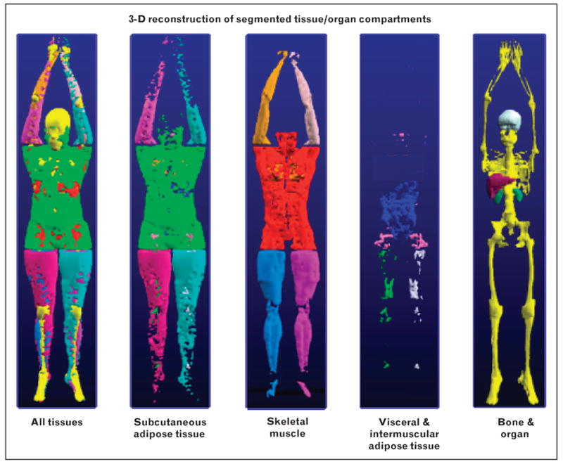
Whole body axial images can be rapidly acquired on most clinical magnetic resonance imaging scanners. The images can then be semi-automatically segmented into subcutaneous, visceral, and intramuscular adipose tissue. Specific pediatric studies of errors related to slice gap and number are available. The acquisition of scans in healthy and premature infants is now feasible with recent technological advances. Spectroscopic, Dixon, and other approaches can be used to quantify the lipid content of liver, skeletal muscle, and other organs. Protocol selection is based on factors such as subject age and cost. Particular attention should be directed towards identification of landmarks in growth studies. Recent advances promise to reduce the requirement of subjects to remain motionless for relatively long periods.
Magnetic resonance imaging and spectroscopy are safe, practical, and widely available methods for phenotyping adiposity in children that open new opportunities for metabolism and nutritional research.
为了在体内表征肥胖儿童病情的表型、严重程度和治疗效果,需要准确测量其肥胖程度。磁共振成像和波谱学具有非侵入性且安全的特点,为表征儿童肥胖的关键方面提供了一种重要的新方法。本综述聚焦于非侵入性磁共振成像和波谱学在量化儿童全身及局部肥胖、绘制脂肪组织分布图以及评估特定代谢紊乱方面的最新进展。目的是为研究儿童身体成分和代谢的研究人员提供一份以磁共振方法为重点的概述。
在大多数临床磁共振成像扫描仪上都可以快速获取全身轴向图像。然后可以将这些图像半自动分割为皮下、内脏和肌肉内脂肪组织。现有针对与切片间隙和数量相关误差的特定儿科研究。随着最近的技术进步,现在对健康婴儿和早产儿进行扫描已可行。波谱学、狄克逊(Dixon)及其他方法可用于量化肝脏、骨骼肌和其他器官的脂质含量。方案选择基于受试者年龄和成本等因素。在生长研究中应特别关注地标识别。最近的进展有望减少受试者长时间保持静止的要求。
磁共振成像和波谱学是用于儿童肥胖表型分析的安全、实用且广泛可用的方法,为代谢和营养研究带来了新机遇。