Dawson T M, Bredt D S, Fotuhi M, Hwang P M, Snyder S H
Johns Hopkins University School of Medicine, Department of Neuroscience, Baltimore, MD 21205.
Proc Natl Acad Sci U S A. 1991 Sep 1;88(17):7797-801. doi: 10.1073/pnas.88.17.7797.
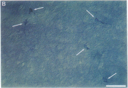
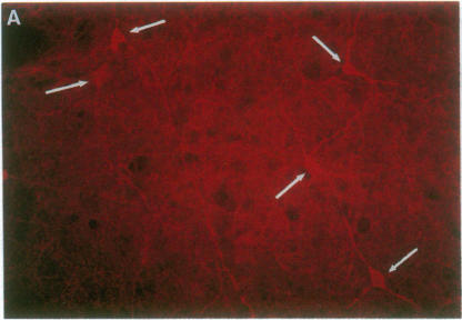
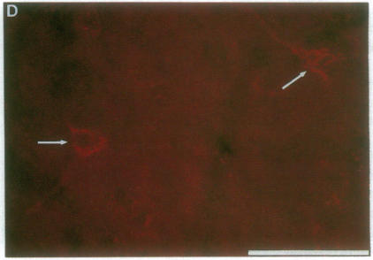
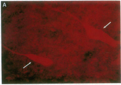
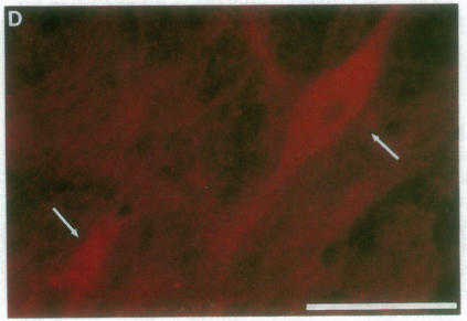
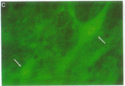
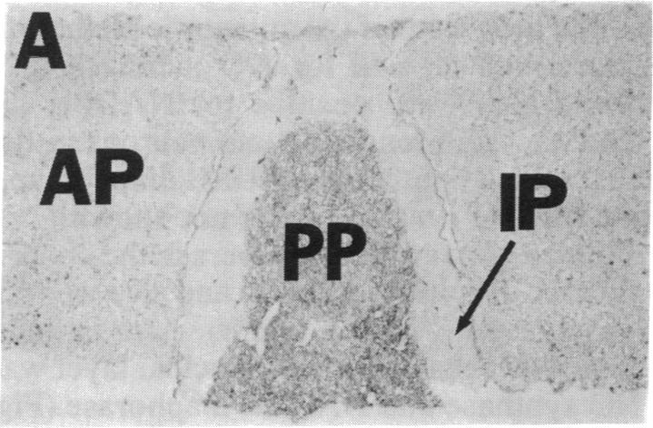
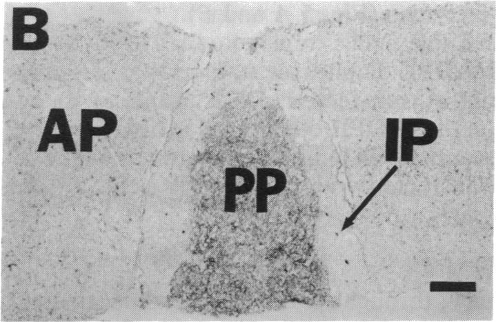
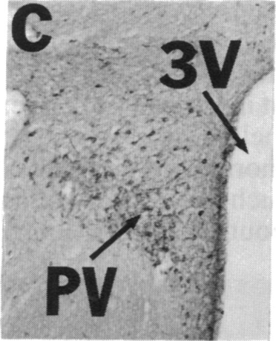
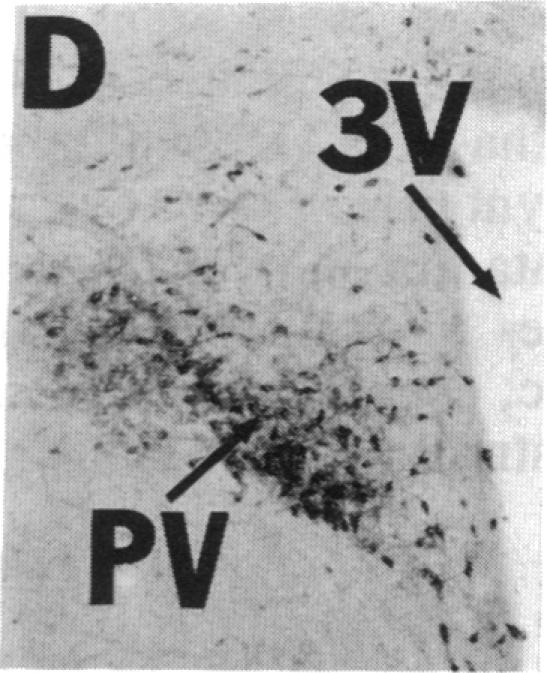
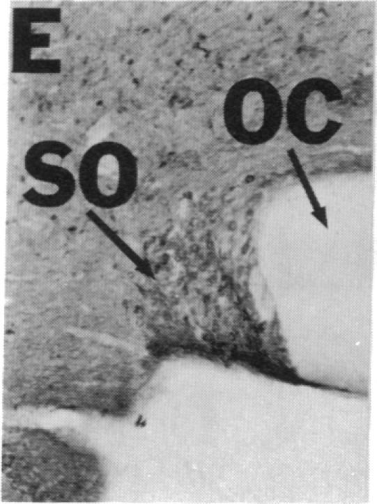
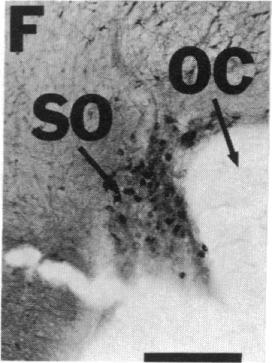
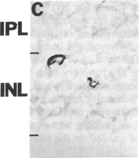
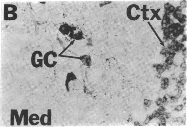
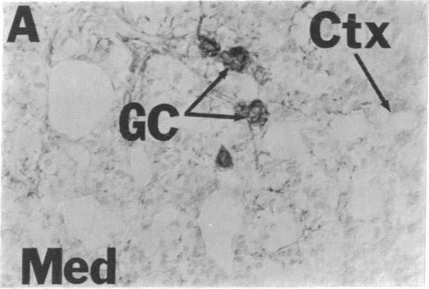
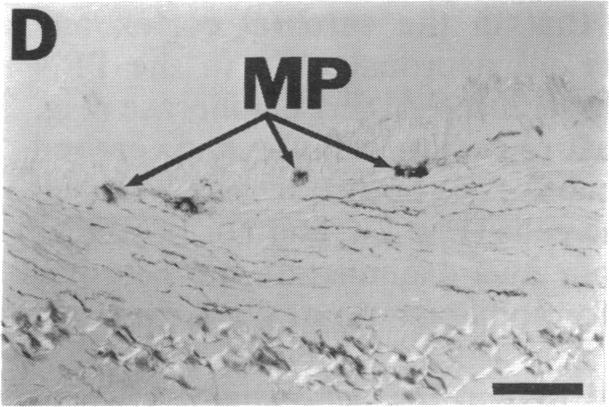
NADPH diaphorase staining neurons, uniquely resistant to toxic insults and neurodegenerative disorders, have been colocalized with neurons in the brain and peripheral tissue containing nitric oxide synthase (EC 1.14.23.-), which generates nitric oxide (NO), a recently identified neuronal messenger molecule. In the corpus striatum and cerebral cortex, NO synthase immunoreactivity and NADPH diaphorase staining are colocalized in medium to large aspiny neurons. These same neurons colocalize with somatostatin and neuropeptide Y immunoreactivity. NO synthase immunoreactivity and NADPH diaphorase staining are colocalized in the pedunculopontine nucleus with choline acetyltransferase-containing cells and are also colocalized in amacrine cells of the inner nuclear layer and ganglion cells of the retina, myenteric plexus neurons of the intestine, and ganglion cells of the adrenal medulla. Transfection of human kidney cells with NO synthase cDNA elicits NADPH diaphorase staining. The ratio of NO synthase to NADPH diaphorase staining in the transfected cells is the same as in neurons, indicating that NO synthase fully accounts for observed NADPH staining. The identity of neuronal NO synthase and NADPH diaphorase suggests a role for NO in modulating neurotoxicity.
烟酰胺腺嘌呤二核苷酸磷酸(NADPH)黄递酶染色神经元对毒性损伤和神经退行性疾病具有独特的抗性,已被证明与大脑和外周组织中含有一氧化氮合酶(EC 1.14.23.-)的神经元共定位,该酶可产生一氧化氮(NO),一种最近被发现的神经元信使分子。在纹状体和大脑皮层中,一氧化氮合酶免疫反应性和NADPH黄递酶染色在中等至大型无棘神经元中共定位。这些相同的神经元与生长抑素和神经肽Y免疫反应性共定位。一氧化氮合酶免疫反应性和NADPH黄递酶染色在脚桥核中与含胆碱乙酰转移酶的细胞共定位,也在视网膜内核层的无长突细胞、视网膜神经节细胞、肠肌间神经丛神经元以及肾上腺髓质神经节细胞中共定位。用一氧化氮合酶cDNA转染人肾细胞可引发NADPH黄递酶染色。转染细胞中一氧化氮合酶与NADPH黄递酶染色的比例与神经元中的相同,表明一氧化氮合酶完全解释了观察到的NADPH染色。神经元一氧化氮合酶和NADPH黄递酶的一致性表明NO在调节神经毒性中起作用。