• 文献检索
  • 文档翻译
  • 深度研究
  • 学术资讯
  • Suppr Zotero 插件Zotero 插件
  • 邀请有礼
  • 套餐&价格
  • 历史记录
应用&插件
Suppr Zotero 插件Zotero 插件浏览器插件Mac 客户端Windows 客户端微信小程序
定价
高级版会员购买积分包购买API积分包
服务
文献检索文档翻译深度研究API 文档MCP 服务
关于我们
关于 Suppr公司介绍联系我们用户协议隐私条款
关注我们

Suppr 超能文献

核心技术专利:CN118964589B侵权必究
粤ICP备2023148730 号-1Suppr @ 2026

文献检索

告别复杂PubMed语法,用中文像聊天一样搜索,搜遍4000万医学文献。AI智能推荐,让科研检索更轻松。

立即免费搜索

文件翻译

保留排版,准确专业,支持PDF/Word/PPT等文件格式,支持 12+语言互译。

免费翻译文档

深度研究

AI帮你快速写综述,25分钟生成高质量综述,智能提取关键信息,辅助科研写作。

立即免费体验

一种用于人类部分性癫痫患者脑内电极记录的脑电图信号的生理合理时空模型。

A physiologically plausible spatio-temporal model for EEG signals recorded with intracerebral electrodes in human partial epilepsy.

作者信息

Cosandier-Rimélé Delphine, Badier Jean-Michel, Chauvel Patrick, Wendling Fabrice

机构信息

INSERM, U642, Laboratoire Traitement du Signal et de l'Image, Campus de Beaulieu, Université de Rennes 1, LTSI, 35042, France.

出版信息

IEEE Trans Biomed Eng. 2007 Mar;54(3):380-8. doi: 10.1109/TBME.2006.890489.

DOI:10.1109/TBME.2006.890489
PMID:17355049
原文链接:https://pmc.ncbi.nlm.nih.gov/articles/PMC1978245/
Abstract

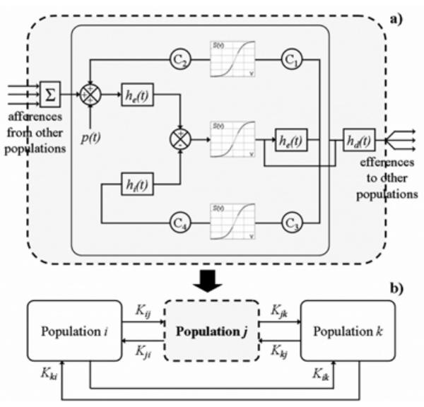
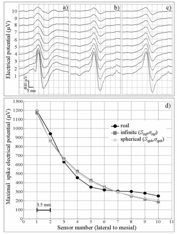
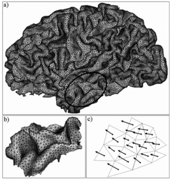
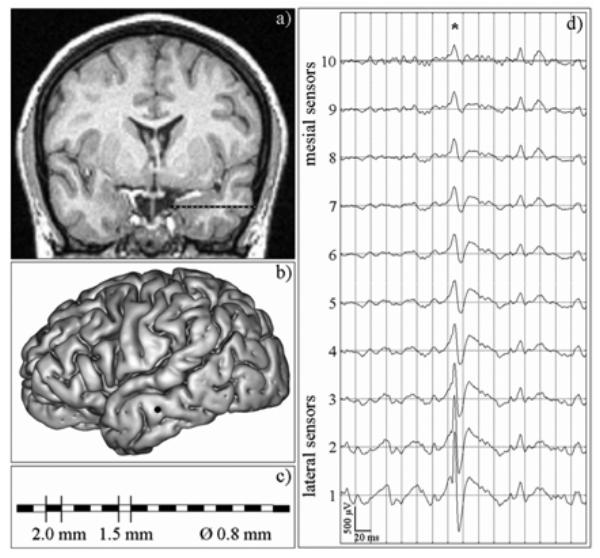
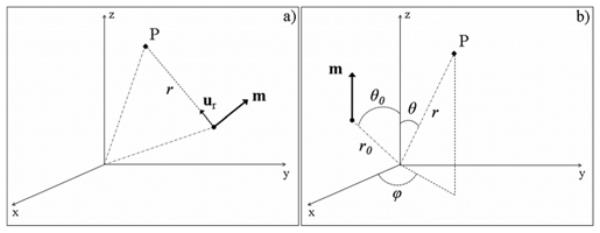
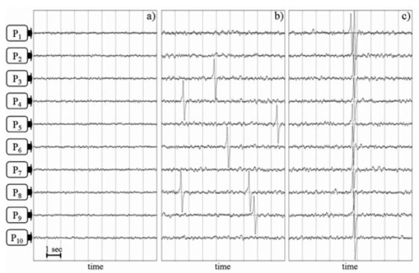
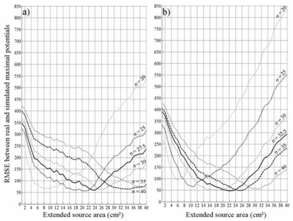
Stereoelectroencephalography (depth-EEG signals) is a presurgical investigation technique of drug-resistant partial epilepsy, in which multiple sensor intracerebral electrodes are used to directly record brain electrical activity. In order to interpret depth-EEG signals, we developed an extended source model which connects two levels of representation: (1) a distributed current dipole model which describes the spatial distribution of neuronal sources; (2) a model of coupled neuronal populations which describes their temporal dynamics. From this extended source model, depth-EEG signals were simulated from the forward solution at each electrode sensor located inside the brain. Results showed that realistic transient epileptiform activities (spikes) are obtained under specific conditions in the model in terms of degree of coupling between neuronal populations and spatial extent of the source. In particular, the cortical area involved in the generation of epileptic spikes was estimated to vary from 18 to 25 cm2, for brain conductivity values ranging from 30 to 35 x 10(-5) S/mm, for high coupling degree between neuronal populations and for a volume conductor model that accounts for the three main tissues of the head (brain, skull, and scalp). This study provides insight into the relationship between spatio-temporal properties of cortical neuronal sources and depth-EEG signals.

摘要

立体脑电图(深度脑电图信号)是一种用于耐药性局灶性癫痫的术前检查技术,该技术使用多个颅内传感器电极直接记录脑电活动。为了解释深度脑电图信号,我们开发了一种扩展源模型,该模型连接了两个表征层次:(1)描述神经元源空间分布的分布式电流偶极子模型;(2)描述神经元群体时间动态的耦合神经元群体模型。基于这个扩展源模型,从位于脑内的每个电极传感器的正向解模拟出深度脑电图信号。结果表明,根据神经元群体之间的耦合程度和源的空间范围,在模型的特定条件下可获得逼真的瞬态癫痫样活动(尖峰)。特别是,对于脑电导率值在30至35×10(-5)S/mm之间、神经元群体之间耦合程度高且考虑头部三个主要组织(脑、颅骨和头皮)的容积导体模型,参与癫痫尖峰产生的皮质面积估计在18至25平方厘米之间变化。本研究深入探讨了皮质神经元源的时空特性与深度脑电图信号之间的关系。

https://cdn.ncbi.nlm.nih.gov/pmc/blobs/0098/1978245/254d541e92e7/halms144531f7.jpg
https://cdn.ncbi.nlm.nih.gov/pmc/blobs/0098/1978245/f07cf95b384f/halms144531f1.jpg
https://cdn.ncbi.nlm.nih.gov/pmc/blobs/0098/1978245/edd7b27f707a/halms144531f2.jpg
https://cdn.ncbi.nlm.nih.gov/pmc/blobs/0098/1978245/a5e3c836b435/halms144531f3.jpg
https://cdn.ncbi.nlm.nih.gov/pmc/blobs/0098/1978245/38b931b6ac8f/halms144531f4.jpg
https://cdn.ncbi.nlm.nih.gov/pmc/blobs/0098/1978245/07323c836e7f/halms144531f5.jpg
https://cdn.ncbi.nlm.nih.gov/pmc/blobs/0098/1978245/613f30b1b1bc/halms144531f6.jpg
https://cdn.ncbi.nlm.nih.gov/pmc/blobs/0098/1978245/254d541e92e7/halms144531f7.jpg

相似文献

1
A physiologically plausible spatio-temporal model for EEG signals recorded with intracerebral electrodes in human partial epilepsy.一种用于人类部分性癫痫患者脑内电极记录的脑电图信号的生理合理时空模型。
IEEE Trans Biomed Eng. 2007 Mar;54(3):380-8. doi: 10.1109/TBME.2006.890489.
2
The neuronal sources of EEG: modeling of simultaneous scalp and intracerebral recordings in epilepsy.脑电图的神经元来源:癫痫患者头皮与脑内同步记录的建模
Neuroimage. 2008 Aug 1;42(1):135-46. doi: 10.1016/j.neuroimage.2008.04.185. Epub 2008 Apr 25.
3
Dipole modeling of epileptic spikes can be accurate or misleading.癫痫棘波的偶极子建模可能准确,也可能产生误导。
Epilepsia. 2005 Mar;46(3):397-408. doi: 10.1111/j.0013-9580.2005.31404.x.
4
Dense array EEG: methodology and new hypothesis on epilepsy syndromes.密集阵列脑电图:癫痫综合征的方法学与新假说
Epilepsia. 2008;49 Suppl 3:3-14. doi: 10.1111/j.1528-1167.2008.01505.x.
5
Computational modeling of epileptic activity: from cortical sources to EEG signals.癫痫活动的计算建模:从皮质源到 EEG 信号。
J Clin Neurophysiol. 2010 Dec;27(6):465-70. doi: 10.1097/WNP.0b013e3182005dcd.
6
Spatio-temporal cortical source imaging of brain electrical activity by means of time-varying parametric projection filter.通过时变参数投影滤波器对脑电活动进行时空皮质源成像。
IEEE Trans Biomed Eng. 2004 May;51(5):768-77. doi: 10.1109/TBME.2004.824142.
7
Expanded head surface EEG electrode array: an application to display the voltage topography of focal epileptiform discharges of mesiotemporal origin.扩展头部表面脑电图电极阵列:用于显示颞叶内侧起源的局灶性癫痫样放电电压地形图的应用。
J Clin Neurophysiol. 1991 Oct;8(4):442-51.
8
Clinical utility of current-generation dipole modelling of scalp EEG.当前一代头皮脑电图偶极子模型的临床应用价值
Clin Neurophysiol. 2007 Nov;118(11):2344-61. doi: 10.1016/j.clinph.2007.08.016. Epub 2007 Sep 21.
9
Source localization of scalp-EEG interictal spikes in posterior cortex epilepsies investigated by HR-EEG and SEEG.通过高分辨率脑电图(HR-EEG)和立体定向脑电图(SEEG)研究后皮质癫痫中头皮脑电图发作间期棘波的来源定位。
Epilepsia. 2009 Feb;50(2):276-89. doi: 10.1111/j.1528-1167.2008.01742.x. Epub 2008 Aug 19.
10
Comparison between realistic and spherical approaches in EEG forward modelling.脑电图正向建模中现实方法与球形方法的比较。
Biomed Tech (Berl). 2010 Jun;55(3):133-46. doi: 10.1515/BMT.2010.010.

引用本文的文献

1
Effects of the spatial resolution of the Virtual Epileptic Patient on the identification of epileptogenic networks.虚拟癫痫患者空间分辨率对致痫网络识别的影响。
Imaging Neurosci (Camb). 2024 May 8;2. doi: 10.1162/imag_a_00153. eCollection 2024.
2
Seizure Sources Can Be Imaged from Scalp EEG by Means of Biophysically Constrained Deep Neural Networks.癫痫发作源可通过生物物理约束深度神经网络从头皮脑电图成像。
Adv Sci (Weinh). 2024 Dec;11(47):e2405246. doi: 10.1002/advs.202405246. Epub 2024 Oct 29.
3
Correspondence between scalp-EEG and stereoelectroencephalography seizure-onset patterns in patients with MRI-negative drug-resistant focal epilepsy.

本文引用的文献

1
A computationally efficient method for accurately solving the EEG forward problem in a finely discretized head model.一种在精细离散头部模型中精确求解脑电正向问题的计算高效方法。
Clin Neurophysiol. 2005 Oct;116(10):2302-14. doi: 10.1016/j.clinph.2005.07.010.
2
Intracranial EEG substrates of scalp EEG interictal spikes.头皮脑电图发作间期棘波的颅内脑电图基质
Epilepsia. 2005 May;46(5):669-76. doi: 10.1111/j.1528-1167.2005.11404.x.
3
Dipole modeling of epileptic spikes can be accurate or misleading.癫痫棘波的偶极子建模可能准确,也可能产生误导。
头皮脑电图与 MRI 阴性耐药性局灶性癫痫患者的立体脑电图发作起始模式的相关性。
Epilepsia Open. 2024 Apr;9(2):568-581. doi: 10.1002/epi4.12886. Epub 2024 Jan 25.
4
Deep learning based source imaging provides strong sublobar localization of epileptogenic zone from MEG interictal spikes.基于深度学习的源成像技术可从 MEG 发作间期棘波中对致痫区进行强有力的亚区定位。
Neuroimage. 2023 Nov 1;281:120366. doi: 10.1016/j.neuroimage.2023.120366. Epub 2023 Sep 15.
5
Electrophysiological Brain Connectivity: Theory and Implementation.脑电生理连接:理论与实现
IEEE Trans Biomed Eng. 2019 May 7. doi: 10.1109/TBME.2019.2913928.
6
A detailed anatomical and mathematical model of the hippocampal formation for the generation of sharp-wave ripples and theta-nested gamma oscillations.用于产生尖波涟漪和theta嵌套伽马振荡的海马结构的详细解剖学和数学模型。
J Comput Neurosci. 2018 Dec;45(3):207-221. doi: 10.1007/s10827-018-0704-x. Epub 2018 Oct 31.
7
Auditory steady state responses and cochlear implants: Modeling the artifact-response mixture in the perspective of denoising.听觉稳态反应与人工耳蜗:从去噪角度看伪迹-反应混合物模型。
PLoS One. 2017 Mar 28;12(3):e0174462. doi: 10.1371/journal.pone.0174462. eCollection 2017.
8
Analysis and Enhancements of a Prolific Macroscopic Model of Epilepsy.一种多产癫痫宏观模型的分析与改进
Scientifica (Cairo). 2016;2016:3628247. doi: 10.1155/2016/3628247. Epub 2016 Apr 7.
9
Intracranial EEG potentials estimated from MEG sources: A new approach to correlate MEG and iEEG data in epilepsy.从脑磁图(MEG)源估计颅内脑电图(EEG)电位:一种关联癫痫中MEG和颅内EEG数据的新方法。
Hum Brain Mapp. 2016 May;37(5):1661-83. doi: 10.1002/hbm.23127. Epub 2016 Mar 2.
10
Analysis of the behavior of a seizure neural mass model using describing functions.使用描述函数对癫痫神经团模型的行为进行分析。
J Med Signals Sens. 2013 Jan;3(1):2-14.
Epilepsia. 2005 Mar;46(3):397-408. doi: 10.1111/j.0013-9580.2005.31404.x.
4
A method to identify reproducible subsets of co-activated structures during interictal spikes. Application to intracerebral EEG in temporal lobe epilepsy.一种在发作间期棘波期间识别共同激活结构的可重复子集的方法。应用于颞叶癫痫的颅内脑电图。
Clin Neurophysiol. 2005 Feb;116(2):443-55. doi: 10.1016/j.clinph.2004.08.010.
5
In vivo measurement of the brain and skull resistivities using an EIT-based method and realistic models for the head.使用基于电阻抗断层成像(EIT)的方法和逼真的头部模型对大脑和颅骨电阻率进行体内测量。
IEEE Trans Biomed Eng. 2003 Jun;50(6):754-67. doi: 10.1109/tbme.2003.812164.
6
Spatiotemporal forward solution of the EEG and MEG using network modeling.使用网络建模的脑电图(EEG)和脑磁图(MEG)时空正向解
IEEE Trans Med Imaging. 2002 May;21(5):493-504. doi: 10.1109/TMI.2002.1009385.
7
Conductivity of living intracranial tissues.
Phys Med Biol. 2001 Jun;46(6):1611-6. doi: 10.1088/0031-9155/46/6/302.
8
The conductivity of the human skull: results of in vivo and in vitro measurements.人类颅骨的电导率:体内和体外测量结果
IEEE Trans Biomed Eng. 2000 Nov;47(11):1487-92. doi: 10.1109/TBME.2000.880100.
9
Relevance of nonlinear lumped-parameter models in the analysis of depth-EEG epileptic signals.非线性集总参数模型在深度脑电图癫痫信号分析中的相关性。
Biol Cybern. 2000 Oct;83(4):367-78. doi: 10.1007/s004220000160.
10
Emerging insights into the genesis of epilepsy.对癫痫起源的新见解。
Nature. 1999 Jun 24;399(6738 Suppl):A15-22. doi: 10.1038/399a015.