• 文献检索
  • 文档翻译
  • 深度研究
  • 学术资讯
  • Suppr Zotero 插件Zotero 插件
  • 邀请有礼
  • 套餐&价格
  • 历史记录
应用&插件
Suppr Zotero 插件Zotero 插件浏览器插件Mac 客户端Windows 客户端微信小程序
定价
高级版会员购买积分包购买API积分包
服务
文献检索文档翻译深度研究API 文档MCP 服务
关于我们
关于 Suppr公司介绍联系我们用户协议隐私条款
关注我们

Suppr 超能文献

核心技术专利:CN118964589B侵权必究
粤ICP备2023148730 号-1Suppr @ 2026

文献检索

告别复杂PubMed语法,用中文像聊天一样搜索,搜遍4000万医学文献。AI智能推荐,让科研检索更轻松。

立即免费搜索

文件翻译

保留排版,准确专业,支持PDF/Word/PPT等文件格式,支持 12+语言互译。

免费翻译文档

深度研究

AI帮你快速写综述,25分钟生成高质量综述,智能提取关键信息,辅助科研写作。

立即免费体验

细菌芽孢的快速诊断薄切片电子显微镜检查

Rapid diagnostic thin section electron microscopy of bacterial endospores.

作者信息

Laue Michael, Niederwöhrmeier Bärbel, Bannert Norbert

机构信息

Centre for Biological Safety 4, Robert Koch Institute, Nordufer 20, D-13353 Berlin, Germany.

出版信息

J Microbiol Methods. 2007 Jul;70(1):45-54. doi: 10.1016/j.mimet.2007.03.006. Epub 2007 Mar 30.

DOI:10.1016/j.mimet.2007.03.006
PMID:17466397
原文链接:https://pmc.ncbi.nlm.nih.gov/articles/PMC7114357/
Abstract

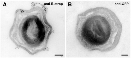
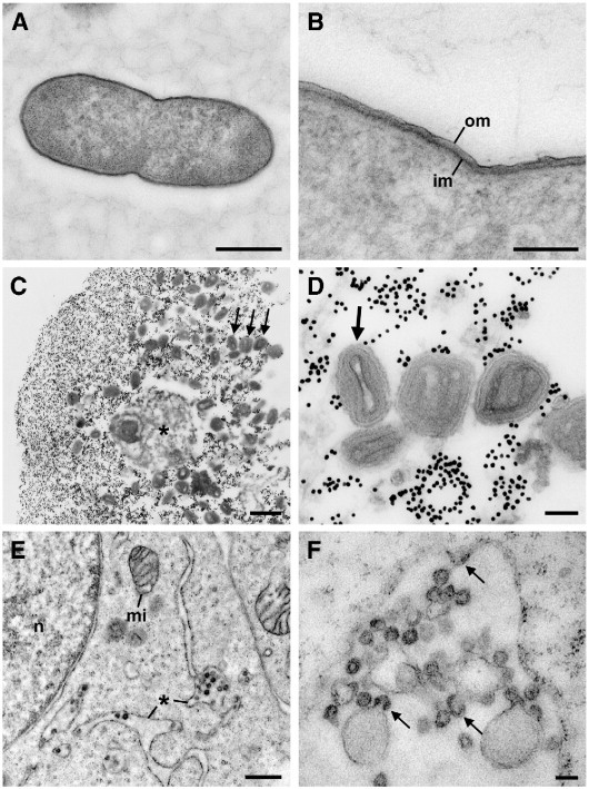
Emerging infectious diseases such as SARS and the bioterror attacks with anthrax spores that occurred after September 11th, 2001 have highlighted the need to be better prepared for the detection and management of infectious pathogens that threaten public health. Negative staining electron microscopy is one method used to screen environmental and clinical samples for relevant infectious pathogens. Unfortunately, bacterial endospores, like those of Bacillus anthracis, are difficult to identify using this method because of their density that prevents imaging of structural details. Thin section electron microscopy would be an alternative method but this usually requires a few days for preparation and diagnosis. In the present paper we describe the development of a rapid thin section protocol, using mainly Bacillus subtilis spores as a model, which allows an unequivocal diagnosis of endospores within 2 h. The protocol involves chemical fixation assisted by heat or microwaves, rapid dehydration, embedding in the low-viscosity resin LR White and chemically enhanced polymerization. Structural preservation of spores is comparable to preservation after standard Epon embedding. Immunolabeling experiments using B. atrophaeus spores and a specific antibody suggest that the protocol preserves significant antigenicity for on-section immunocytochemistry and therefore offers the possibility for the strain typing of spores using specific antibodies. Further experiments with vegetative bacteria, viruses and cell cultures indicate that the rapid thin section protocol not only preserves spores but also other biological structures. Because of its universality and speed the described protocol complements negative staining electron microscopy as a front line method for the morphology-based diagnosis of pathogens in environmental and clinical samples.

摘要

诸如严重急性呼吸综合征(SARS)等新出现的传染病以及2001年9月11日之后发生的炭疽芽孢生物恐怖袭击事件凸显了对威胁公共卫生的传染性病原体的检测和管理做好更充分准备的必要性。负染色电子显微镜检查是用于筛查环境和临床样本中相关传染性病原体的一种方法。不幸的是,像炭疽芽孢杆菌的芽孢那样的细菌芽孢,由于其密度会妨碍结构细节成像,所以难以用这种方法识别。超薄切片电子显微镜检查将是一种替代方法,但这通常需要几天时间来制备和诊断。在本文中,我们描述了一种快速超薄切片方案的开发,主要以枯草芽孢杆菌芽孢为模型,该方案能在2小时内明确诊断芽孢。该方案包括加热或微波辅助的化学固定、快速脱水、包埋于低粘度树脂LR White以及化学增强聚合。芽孢的结构保存与标准环氧树脂包埋后的保存效果相当。使用萎缩芽孢杆菌芽孢和特异性抗体进行的免疫标记实验表明,该方案为切片免疫细胞化学保留了显著的抗原性,因此提供了使用特异性抗体对芽孢进行菌株分型的可能性。对营养细菌、病毒和细胞培养物进行的进一步实验表明,快速超薄切片方案不仅能保存芽孢,还能保存其他生物结构。由于其通用性和速度,所描述的方案可作为负染色电子显微镜检查的补充,作为对环境和临床样本中病原体进行基于形态学诊断的一线方法。

https://cdn.ncbi.nlm.nih.gov/pmc/blobs/4928/7114357/268e15760636/gr6.jpg
https://cdn.ncbi.nlm.nih.gov/pmc/blobs/4928/7114357/2c60c2fda53f/gr1.jpg
https://cdn.ncbi.nlm.nih.gov/pmc/blobs/4928/7114357/2b8edd9b90ac/gr2.jpg
https://cdn.ncbi.nlm.nih.gov/pmc/blobs/4928/7114357/a1036e6a33e4/gr3.jpg
https://cdn.ncbi.nlm.nih.gov/pmc/blobs/4928/7114357/7c483d15ef26/gr4.jpg
https://cdn.ncbi.nlm.nih.gov/pmc/blobs/4928/7114357/e805f39bb5e0/gr5.jpg
https://cdn.ncbi.nlm.nih.gov/pmc/blobs/4928/7114357/268e15760636/gr6.jpg

相似文献

1
Rapid diagnostic thin section electron microscopy of bacterial endospores.细菌芽孢的快速诊断薄切片电子显微镜检查
J Microbiol Methods. 2007 Jul;70(1):45-54. doi: 10.1016/j.mimet.2007.03.006. Epub 2007 Mar 30.
2
Rapid and reliable detection of bacterial endospores in environmental samples by diagnostic electron microscopy combined with X-ray microanalysis.利用诊断电子显微镜结合 X 射线微分析快速可靠地检测环境样品中的细菌芽孢。
J Microbiol Methods. 2013 Jul;94(1):13-21. doi: 10.1016/j.mimet.2013.03.026. Epub 2013 Apr 17.
3
Dormant Bacillus spores protect their DNA in crystalline nucleoids against environmental stress.休眠的芽孢杆菌孢子在晶体类核中保护其DNA免受环境压力的影响。
J Struct Biol. 2015 Aug;191(2):156-64. doi: 10.1016/j.jsb.2015.06.019. Epub 2015 Jun 19.
4
Combined scanning transmission X-ray and electron microscopy for the characterization of bacterial endospores.结合扫描透射X射线和电子显微镜对细菌芽孢进行表征
FEMS Microbiol Lett. 2014 Sep;358(2):188-93. doi: 10.1111/1574-6968.12539. Epub 2014 Aug 12.
5
Postembedding immunolabeling of thin sections of Drosophila tissues for transmission electron microscopy.用于透射电子显微镜的果蝇组织薄切片的包埋后免疫标记。
Cold Spring Harb Protoc. 2012 Jul 1;2012(7):810-2. doi: 10.1101/pdb.prot068437.
6
Wet and dry density of Bacillus anthracis and other Bacillus species.炭疽芽孢杆菌及其他芽孢杆菌属的湿密度和干密度。
J Appl Microbiol. 2008 Jul;105(1):68-77. doi: 10.1111/j.1365-2672.2008.03758.x. Epub 2008 Feb 20.
7
Novel technology for rapid species-specific detection of Bacillus spores.用于快速进行芽孢杆菌孢子物种特异性检测的新技术。
Biomol Eng. 2006 Jun;23(2-3):119-27. doi: 10.1016/j.bioeng.2005.12.003. Epub 2006 Feb 23.
8
Inactivation and ultrastructure analysis of Bacillus spp. and Clostridium perfringens spores.芽孢杆菌属和产气荚膜梭菌孢子的灭活及超微结构分析
Microsc Microanal. 2014 Feb;20(1):238-44. doi: 10.1017/S1431927613013949.
9
Electron microscopy of spores of Bacillus megaterium with special reference to the effects of fixation and thin sectioning.巨大芽孢杆菌孢子的电子显微镜观察,特别提及固定和超薄切片的影响。
J Cell Biol. 1962 Jun;13(3):423-35. doi: 10.1083/jcb.13.3.423.
10
Tetrodotoxin-producing Bacillus sp. from the ribbon worm (Nemertea) Cephalothrix simula (Iwata, 1952).来自纽形动物(Nemertea)拟头状纽虫(Cephalothrix simula,岩田,1952)的产河豚毒素芽孢杆菌属细菌
Toxicon. 2014 Jul;85:46-51. doi: 10.1016/j.toxicon.2014.04.015. Epub 2014 May 9.

引用本文的文献

1
Diagnostic electron microscopy in human infectious diseases - Methods and applications.人类传染病诊断电子显微镜检查——方法与应用
J Microsc. 2025 Sep;299(3):186-205. doi: 10.1111/jmi.13370. Epub 2024 Nov 19.
2
Visualization of SARS-CoV-2 particles in naso/oropharyngeal swabs by thin section electron microscopy.通过超薄切片电子显微镜观察鼻咽拭子中的 SARS-CoV-2 病毒颗粒。
Virol J. 2023 Feb 6;20(1):21. doi: 10.1186/s12985-023-01981-9.
3
Antibacterial activity of geraniin from sugar maple leaves: an ultrastructural study with the phytopathogen pv. .

本文引用的文献

1
Spores of Bacillus subtilis: their resistance to and killing by radiation, heat and chemicals.枯草芽孢杆菌的孢子:它们对辐射、热和化学物质的抗性及被其杀灭的情况。
J Appl Microbiol. 2006 Sep;101(3):514-25. doi: 10.1111/j.1365-2672.2005.02736.x.
2
Microwave-assisted tissue processing for same-day EM-diagnosis of potential bioterrorism and clinical samples.用于潜在生物恐怖主义和临床样本当日电镜诊断的微波辅助组织处理
Micron. 2006;37(6):577-90. doi: 10.1016/j.micron.2005.11.015.
3
Arguments pro disinfection in diagnostic electron microscopy: a response to Madeley and Biel.
糖枫叶中老鹳草素的抗菌活性:对植物病原体pv. 的超微结构研究
J Plant Pathol. 2021;103(2):461-471. doi: 10.1007/s42161-021-00743-2. Epub 2021 Feb 3.
4
Rapid Viral Diagnosis of Orthopoxviruses by Electron Microscopy: Optional or a Must?电子显微镜在正痘病毒快速诊断中的应用:是可选还是必需?
Viruses. 2018 Mar 22;10(4):142. doi: 10.3390/v10040142.
5
Detection limit of negative staining electron microscopy for the diagnosis of bioterrorism-related micro-organisms.负染电子显微镜检测下限对生物恐怖相关微生物诊断的影响。
J Appl Microbiol. 2010 Oct;109(4):1159-68. doi: 10.1111/j.1365-2672.2010.04737.x. Epub 2010 Aug 19.
支持诊断电子显微镜消毒的论据:对马德利和比尔的回应。
J Infect. 2007 Mar;54(3):307-8; author reply 308-9. doi: 10.1016/j.jinf.2006.03.032. Epub 2006 May 22.
4
Application of transmission electron microscopy to the clinical study of viral and bacterial infections: present and future.透射电子显微镜在病毒和细菌感染临床研究中的应用:现状与未来
Micron. 2006;37(2):91-106. doi: 10.1016/j.micron.2005.10.001. Epub 2005 Nov 2.
5
Improvements in epoxy resin embedding methods.环氧树脂包埋方法的改进。
J Biophys Biochem Cytol. 1961 Feb;9(2):409-14. doi: 10.1083/jcb.9.2.409.
6
Microwave and digital imaging technology reduce turnaround times for diagnostic electron microscopy.微波和数字成像技术缩短了诊断电子显微镜检查的周转时间。
Ultrastruct Pathol. 2003 May-Jun;27(3):187-96. doi: 10.1080/01913120309937.
7
Bioterrorism and electron microscopic differentiation of poxviruses from herpesviruses: dos and don'ts.
Ultrastruct Pathol. 2003 May-Jun;27(3):133-40. doi: 10.1080/01913120309932.
8
A multicentre collaboration to investigate the cause of severe acute respiratory syndrome.一项调查严重急性呼吸综合征病因的多中心合作研究。
Lancet. 2003 May 17;361(9370):1730-3. doi: 10.1016/s0140-6736(03)13376-4.
9
Electron microscopy for rapid diagnosis of infectious agents in emergent situations.电子显微镜用于紧急情况下传染病原体的快速诊断。
Emerg Infect Dis. 2003 Mar;9(3):294-303. doi: 10.3201/eid0903.020327.
10
Bacillus subtilis spore coat.枯草芽孢杆菌芽孢衣
Microbiol Mol Biol Rev. 1999 Mar;63(1):1-20. doi: 10.1128/MMBR.63.1.1-20.1999.