Ma P C, Tretiakova M S, Nallasura V, Jagadeeswaran R, Husain A N, Salgia R
Division of Hematology/Oncology, Department of Medicine, University Hospitals of Case Medical Center and Ireland Cancer Center, Case Western Reserve University, Case Comprehensive Cancer Center, Cleveland, OH 44106, USA.
Br J Cancer. 2007 Aug 6;97(3):368-77. doi: 10.1038/sj.bjc.6603884. Epub 2007 Jul 31.
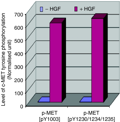
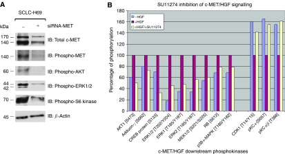
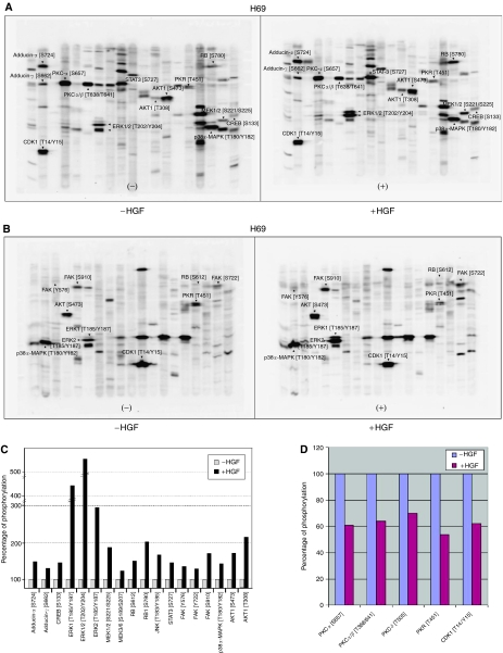
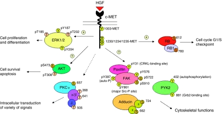
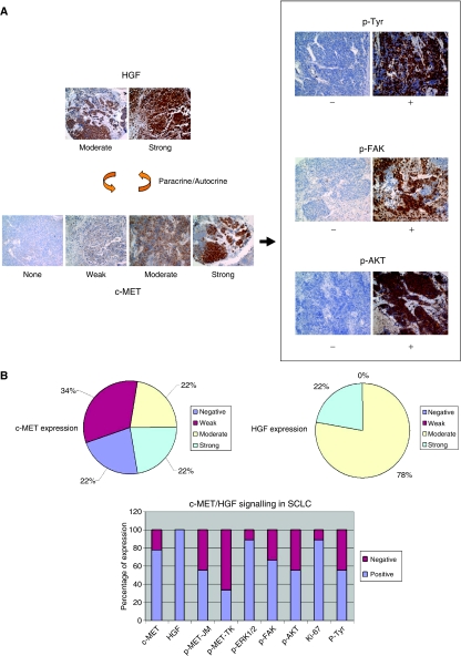
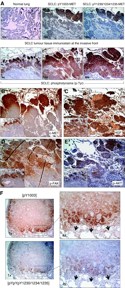
The c-MET receptor can be overexpressed, amplified, or mutated in solid tumours including small cell lung cancer (SCLC). In c-MET-overexpressing SCLC cell line NCI-H69, hepatocyte growth factor (HGF) dramatically induced c-MET phosphorylation at phosphoepitopes pY1230/1234/1235 (catalytic tyrosine kinase), pY1003 (juxtamembrane), and also of paxillin at pY31 (CRKL-binding site). We utilised a global proteomics phosphoantibody array approach to identify further c-MET/HGF signal transduction intermediates in SCLC. Strong HGF induction of specific phosphorylation sites in phosphoproteins involved in c-MET/HGF signal transduction was detected, namely adducin-alpha [S724], adducin-gamma [S662], CREB [S133], ERK1 [T185/Y187], ERK1/2 [T202/Y204], ERK2 [T185/Y187], MAPKK (MEK) 1/2 [S221/S225], MAPKK (MEK) 3/6 [S189/S207], RB [S612], RB1 [S780], JNK [T183/Y185], STAT3 [S727], focal adhesion kinase (FAK) [Y576/S722/S910], p38alpha-MAPK [T180/Y182], and AKT1[S473] and [T308]. Conversely, inhibition of phosphorylation by HGF in protein kinase C (PKC), protein kinase R (PKR), and also CDK1 was identified. Phosphoantibody-based immunohistochemical analysis of SCLC tumour tissue and microarray established the role of c-MET in SCLC biology. This supports a role of c-MET activation in tumour invasive front in the tumour progression and invasion involving FAK and AKT downstream. The c-MET serves as an attractive therapeutic target in SCLC, as shown through small interfering RNA (siRNA) and selective prototype c-MET inhibitor SU11274, inhibiting the phosphorylation of c-MET itself and its downstream molecules such as AKT, S6 kinase, and ERK1/2. Investigation of mechanisms of invasion and, ultimately, metastasis in SCLC would be very useful with these signal transduction molecules.
c-MET受体在包括小细胞肺癌(SCLC)在内的实体瘤中可能会过度表达、扩增或发生突变。在c-MET过表达的SCLC细胞系NCI-H69中,肝细胞生长因子(HGF)显著诱导c-MET在磷酸表位pY1230/1234/1235(催化性酪氨酸激酶)、pY1003(近膜区)以及桩蛋白在pY31(CRKL结合位点)处发生磷酸化。我们利用一种全局蛋白质组学磷酸抗体阵列方法来鉴定SCLC中更多的c-MET/HGF信号转导中间体。检测到HGF对参与c-MET/HGF信号转导的磷蛋白中特定磷酸化位点有强烈诱导作用,即内收蛋白-α [S724]、内收蛋白-γ [S662]、CREB [S133]、ERK1 [T185/Y187]、ERK1/2 [T202/Y204]、ERK2 [T185/Y187]、丝裂原活化蛋白激酶激酶(MEK)1/2 [S221/S225]、MEK 3/6 [S189/S207]、RB [S612]、RB1 [S780]、JNK [T183/Y185]、信号转导子和转录激活子3(STAT3)[S727]、粘着斑激酶(FAK)[Y576/S722/S910]、p38α-丝裂原活化蛋白激酶 [T180/Y182]以及AKT1 [S473]和[T308]。相反,还鉴定出HGF对蛋白激酶C(PKC)、蛋白激酶R(PKR)以及细胞周期蛋白依赖性激酶1(CDK1)的磷酸化有抑制作用。基于磷酸抗体的SCLC肿瘤组织免疫组织化学分析和微阵列确定了c-MET在SCLC生物学中的作用。这支持了c-MET激活在肿瘤进展和侵袭中肿瘤侵袭前沿的作用,涉及下游的FAK和AKT。如通过小干扰RNA(siRNA)和选择性原型c-MET抑制剂SU11274所示,c-MET在SCLC中是一个有吸引力的治疗靶点,其抑制c-MET自身及其下游分子如AKT、S6激酶和ERK1/2的磷酸化。利用这些信号转导分子来研究SCLC的侵袭机制以及最终的转移机制将非常有用。