• 文献检索
  • 文档翻译
  • 深度研究
  • 学术资讯
  • Suppr Zotero 插件Zotero 插件
  • 邀请有礼
  • 套餐&价格
  • 历史记录
应用&插件
Suppr Zotero 插件Zotero 插件浏览器插件Mac 客户端Windows 客户端微信小程序
定价
高级版会员购买积分包购买API积分包
服务
文献检索文档翻译深度研究API 文档MCP 服务
关于我们
关于 Suppr公司介绍联系我们用户协议隐私条款
关注我们

Suppr 超能文献

核心技术专利:CN118964589B侵权必究
粤ICP备2023148730 号-1Suppr @ 2026

文献检索

告别复杂PubMed语法,用中文像聊天一样搜索,搜遍4000万医学文献。AI智能推荐,让科研检索更轻松。

立即免费搜索

文件翻译

保留排版,准确专业,支持PDF/Word/PPT等文件格式,支持 12+语言互译。

免费翻译文档

深度研究

AI帮你快速写综述,25分钟生成高质量综述,智能提取关键信息,辅助科研写作。

立即免费体验

沙粒病毒的形态学与形态发生

Morphology and morphogenesis of arenaviruses.

作者信息

Murphy F A, Whitfield S G

出版信息

Bull World Health Organ. 1975;52(4-6):409-19.

PMID:182396
原文链接:https://pmc.ncbi.nlm.nih.gov/articles/PMC2366645/
Abstract

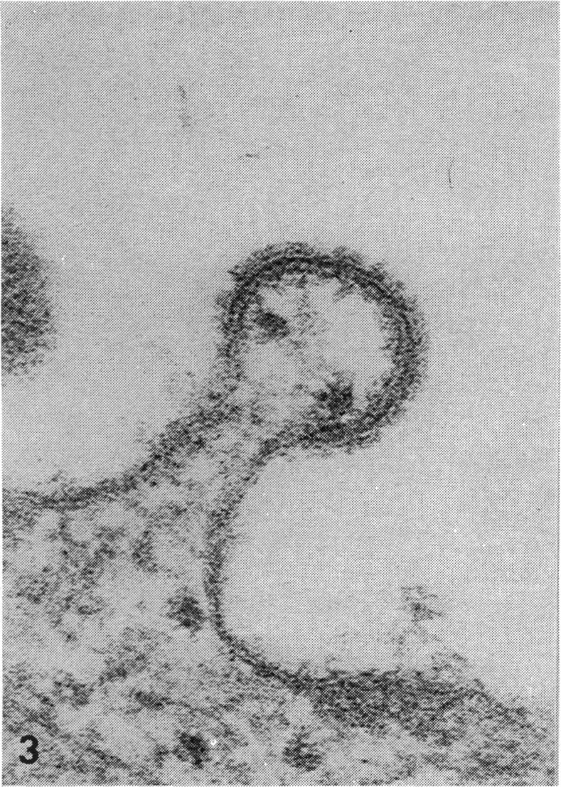
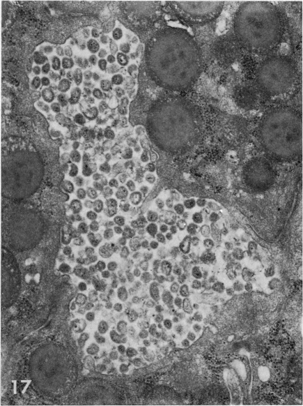
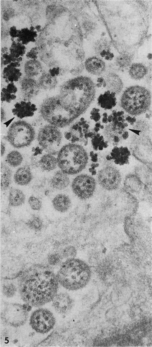
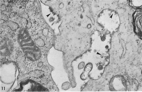
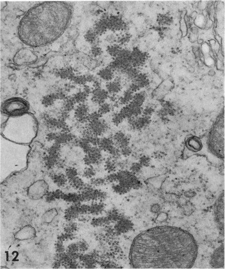
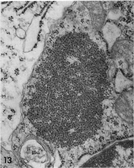
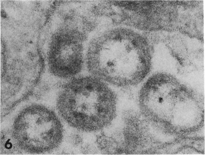
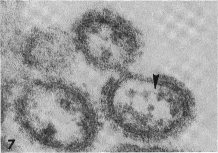
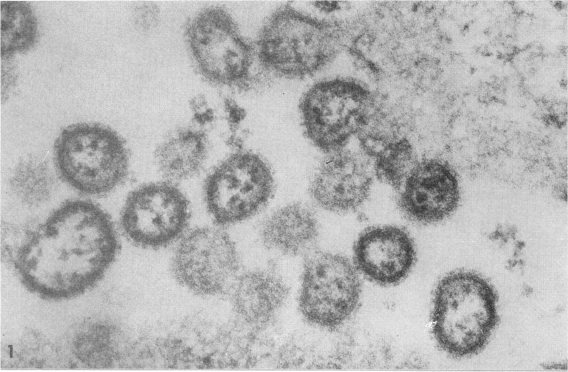
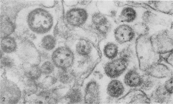
Arenaviruses have unique structural characteristics; they are pleomorphic, have a mean diameter of 110-130 nm, and consist of a membranous envelope with surface projections surrounding an interior containing ribosomes and filaments. Virus particles bud from plasma membranes of infected cells and in many cases large intracytoplasmic inclusion bodies are formed. These characteristics allow generic identification, but not differentiation of individual viruses. Ultrastructural identification of virus particles and pathological processes in infected tissues of man and experimental animals is important in understanding the nature of arenaviral pathogenesis Such identification also contributes to our understanding of the mechanisms of viral shedding and transmission in reservoir host species.

摘要

沙粒病毒具有独特的结构特征;它们形态多样,平均直径为110 - 130纳米,由一个带有表面突起的膜状包膜组成,包膜围绕着一个包含核糖体和细丝的内部结构。病毒粒子从受感染细胞的质膜出芽,在许多情况下会形成大的胞质内包涵体。这些特征有助于进行属的鉴定,但无法区分个别病毒。对人和实验动物感染组织中的病毒粒子和病理过程进行超微结构鉴定,对于理解沙粒病毒发病机制的本质很重要。这种鉴定也有助于我们了解病毒在储存宿主物种中的释放和传播机制。

https://cdn.ncbi.nlm.nih.gov/pmc/blobs/3949/2366645/043c64487046/bullwho00466-0036-d.jpg
https://cdn.ncbi.nlm.nih.gov/pmc/blobs/3949/2366645/5e36ee19591b/bullwho00466-0033-a.jpg
https://cdn.ncbi.nlm.nih.gov/pmc/blobs/3949/2366645/f2199dbb05f9/bullwho00466-0033-b.jpg
https://cdn.ncbi.nlm.nih.gov/pmc/blobs/3949/2366645/b4333d4a1eb7/bullwho00466-0033-c.jpg
https://cdn.ncbi.nlm.nih.gov/pmc/blobs/3949/2366645/9025f7971829/bullwho00466-0035-a.jpg
https://cdn.ncbi.nlm.nih.gov/pmc/blobs/3949/2366645/84509ca318de/bullwho00466-0035-b.jpg
https://cdn.ncbi.nlm.nih.gov/pmc/blobs/3949/2366645/04cc04c3da40/bullwho00466-0035-c.jpg
https://cdn.ncbi.nlm.nih.gov/pmc/blobs/3949/2366645/b8104444c6f9/bullwho00466-0034-a.jpg
https://cdn.ncbi.nlm.nih.gov/pmc/blobs/3949/2366645/2a5da9e3315b/bullwho00466-0034-b.jpg
https://cdn.ncbi.nlm.nih.gov/pmc/blobs/3949/2366645/ff5b8f449f76/bullwho00466-0034-c.jpg
https://cdn.ncbi.nlm.nih.gov/pmc/blobs/3949/2366645/14052fa5bb9d/bullwho00466-0034-d.jpg
https://cdn.ncbi.nlm.nih.gov/pmc/blobs/3949/2366645/6005d7b38319/bullwho00466-0034-e.jpg
https://cdn.ncbi.nlm.nih.gov/pmc/blobs/3949/2366645/a34abece2c83/bullwho00466-0032-a.jpg
https://cdn.ncbi.nlm.nih.gov/pmc/blobs/3949/2366645/7946ad36ee3a/bullwho00466-0032-b.jpg
https://cdn.ncbi.nlm.nih.gov/pmc/blobs/3949/2366645/92638ea7e16e/bullwho00466-0036-a.jpg
https://cdn.ncbi.nlm.nih.gov/pmc/blobs/3949/2366645/4d17d9d07ec9/bullwho00466-0036-b.jpg
https://cdn.ncbi.nlm.nih.gov/pmc/blobs/3949/2366645/dec05fce9c46/bullwho00466-0036-c.jpg
https://cdn.ncbi.nlm.nih.gov/pmc/blobs/3949/2366645/043c64487046/bullwho00466-0036-d.jpg

相似文献

1
Morphology and morphogenesis of arenaviruses.沙粒病毒的形态学与形态发生
Bull World Health Organ. 1975;52(4-6):409-19.
2
Antigenic properties of the arenaviruses.沙粒病毒的抗原特性。
Bull World Health Organ. 1975;52(4-6):421-7.
3
Biochemical and biophysical properties of the arenaviruses.沙粒病毒的生化和生物物理特性。
Prog Med Virol. 1974;18(0):64-80.
4
Arenaviruses.沙粒病毒属
Yale J Biol Med. 1975 May;48(2):115-40.
5
The structure of rodent faunas associated with arenaviral infections.与沙粒病毒感染相关的啮齿动物动物区系结构。
Bull World Health Organ. 1975;52(4-6):621-7.
6
Arenavirus taxonomy: a review.沙粒病毒分类学:综述
Bull World Health Organ. 1975;52(4-6):389-91.
7
Structural insight into arenavirus replication machinery.结构洞察沙粒病毒复制机制。
Nature. 2020 Mar;579(7800):615-619. doi: 10.1038/s41586-020-2114-2. Epub 2020 Mar 18.
8
Sedimentation analysis of the RNAs isolated from interfering particles of Lassa and Machupo viruses.从拉沙病毒和马丘波病毒干扰颗粒中分离出的RNA的沉降分析。
Acta Virol. 1985 Dec;29(6):455-60.
9
Ultrastructure of the replication site in Taura syndrome virus (TSV)-infected cells.感染桃拉综合征病毒(TSV)的细胞中复制位点的超微结构。
Dis Aquat Organ. 2006 Dec 14;73(2):89-101. doi: 10.3354/dao073089.
10
Status of arenavirus vaccines and their application.沙粒病毒疫苗的现状及其应用。
Bull World Health Organ. 1975;52(4-6):729-35.

引用本文的文献

1
The underlying mechanisms of arenaviral entry through matriglycan.沙粒病毒通过基质聚糖进入细胞的潜在机制。
Front Mol Biosci. 2024 Mar 7;11:1371551. doi: 10.3389/fmolb.2024.1371551. eCollection 2024.
2
Lassa Virus Structural Biology and Replication.拉沙病毒结构生物学与复制。
Curr Top Microbiol Immunol. 2023;440:147-164. doi: 10.1007/82_2023_262.
3
How Does Temperature Affect the Dynamics of SARS-CoV-2 M Proteins? Insights from Molecular Dynamics Simulations.温度如何影响 SARS-CoV-2 M 蛋白的动力学?分子动力学模拟的见解。

本文引用的文献

1
Arenoviruses in Vero cells: ultrastructural studies.沙粒病毒在非洲绿猴肾细胞中的超微结构研究。
J Virol. 1970 Oct;6(4):507-18. doi: 10.1128/JVI.6.4.507-518.1970.
2
Lassa fever, a new virus disease of man from West Africa. 3. Isolation and characterization of the virus.拉沙热,一种源自西非的人类新型病毒疾病。3. 病毒的分离与特性鉴定。
Am J Trop Med Hyg. 1970 Jul;19(4):680-91. doi: 10.4269/ajtmh.1970.19.680.
3
Arenoviruses: proposed name for a newly defined virus group.沙粒病毒:一个新定义病毒群的拟命名。
J Membr Biol. 2022 Jun;255(2-3):341-356. doi: 10.1007/s00232-022-00244-y. Epub 2022 May 13.
4
Control of Capsid Transformations during Reovirus Entry.控制呼肠孤病毒进入时的衣壳转换。
Viruses. 2021 Jan 21;13(2):153. doi: 10.3390/v13020153.
5
References.参考文献。
Perspect Med Virol. 1986;2:209-245. doi: 10.1016/S0168-7069(08)70043-0. Epub 2008 May 29.
6
Multifunctional nature of the arenavirus RING finger protein Z.沙粒病毒 RING 指蛋白 Z 的多功能特性。
Viruses. 2012 Nov 9;4(11):2973-3011. doi: 10.3390/v4112973.
7
Lymphocytic choriomeningitis virus. IV. Electron microscopic investigation of the virion.淋巴细胞性脉络丛脑膜炎病毒。IV. 病毒粒子的电子显微镜研究。
Arch Virol. 1983;75(4):229-42. doi: 10.1007/BF01314889.
8
Diagnostic virology using electron microscopic techniques.运用电子显微镜技术的诊断病毒学
Adv Virus Res. 1982;27:1-69. doi: 10.1016/s0065-3527(08)60432-7.
9
Viral contamination of a subline of Toxoplasma gondii RH.弓形虫RH株一个亚系的病毒污染
Infect Immun. 1985 Dec;50(3):917-8. doi: 10.1128/iai.50.3.917-918.1985.
10
Ribonucleoprotein complexes associated with virions of Pichinde virus and Pichinde virus-infected cells.
Med Microbiol Immunol. 1986;175(2-3):79-83. doi: 10.1007/BF02122420.
J Virol. 1970 May;5(5):651-2. doi: 10.1128/JVI.5.5.651-652.1970.
4
Morphological and cytochemical studies on lymphocytic choriomeningitis virus.淋巴细胞性脉络丛脑膜炎病毒的形态学和细胞化学研究
J Virol. 1968 Dec;2(12):1465-78. doi: 10.1128/JVI.2.12.1465-1478.1968.
5
Morphological comparison of Machupo with lymphocytic choriomeningitis virus: basis for a new taxonomic group.马丘波病毒与淋巴细胞性脉络丛脑膜炎病毒的形态学比较:一个新分类群的基础
J Virol. 1969 Oct;4(4):535-41. doi: 10.1128/JVI.4.4.535-541.1969.
6
Use of enzyme-labeled antibody for electron microscope localization of lymphocytic choriomeningitis virus antigens in infected cell cultures.酶标抗体在电子显微镜下对感染细胞培养物中淋巴细胞性脉络丛脑膜炎病毒抗原的定位应用。
J Natl Cancer Inst. 1969 Mar;42(3):497-515.
7
Ultrastructure of Junín virus in mouse whole brain and mouse brain tissue cultures.胡宁病毒在小鼠全脑及小鼠脑组织培养物中的超微结构
J Virol. 1974 Oct;14(4):965-74. doi: 10.1128/JVI.14.4.965-974.1974.
8
Lymphocytic choriomeningitis: ultrastructural pathology.淋巴细胞性脉络丛脑膜炎:超微结构病理学
Exp Mol Pathol. 1975 Oct;23(2):245-65. doi: 10.1016/0014-4800(75)90022-2.
9
The pathology of human Lassa fever.人类拉沙热的病理学
Bull World Health Organ. 1975;52(4-6):535-45.
10
Arenavirus taxonomy: a review.沙粒病毒分类学:综述
Bull World Health Organ. 1975;52(4-6):389-91.