• 文献检索
  • 文档翻译
  • 深度研究
  • 学术资讯
  • Suppr Zotero 插件Zotero 插件
  • 邀请有礼
  • 套餐&价格
  • 历史记录
应用&插件
Suppr Zotero 插件Zotero 插件浏览器插件Mac 客户端Windows 客户端微信小程序
定价
高级版会员购买积分包购买API积分包
服务
文献检索文档翻译深度研究API 文档MCP 服务
关于我们
关于 Suppr公司介绍联系我们用户协议隐私条款
关注我们

Suppr 超能文献

核心技术专利:CN118964589B侵权必究
粤ICP备2023148730 号-1Suppr @ 2026

文献检索

告别复杂PubMed语法,用中文像聊天一样搜索,搜遍4000万医学文献。AI智能推荐,让科研检索更轻松。

立即免费搜索

文件翻译

保留排版,准确专业,支持PDF/Word/PPT等文件格式,支持 12+语言互译。

免费翻译文档

深度研究

AI帮你快速写综述,25分钟生成高质量综述,智能提取关键信息,辅助科研写作。

立即免费体验

立克次氏体与宿主细胞相互作用的体外研究:细胞内生长周期中的延迟期与感染的普氏立克次氏体生长阶段的关系,以及关于宿主细胞碎片抑制立克次氏体摄取的初步观察。

In vitro studies on Rickettsia-host cell interactions: lag phase in intracellular growth cycle as a function of stage of growth of infecting Rickettsia prowazeki, with preliminary observations on inhibition of rickettsial uptake by host cell fragments.

作者信息

Wisseman C L, Waddell A D, Silverman D J

出版信息

Infect Immun. 1976 Jun;13(6):1749-60. doi: 10.1128/iai.13.6.1749-1760.1976.

DOI:10.1128/iai.13.6.1749-1760.1976
PMID:184046
原文链接:https://pmc.ncbi.nlm.nih.gov/articles/PMC420828/
Abstract

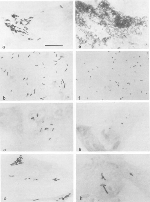
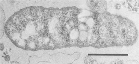
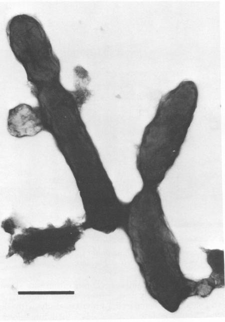
Two Rickettsia prowazeki seeds, an "early" seed in the logarithmic or exponential growth phase and a "late" seed in the stationary or possibly early decline phase, were prepared in chicken embryo (CE) cell cultures and compared with respect to morphology and infection cycle in CE cells in culture. Differences in size and ultrastructure of the organisms in the two seeds were similar to those seen in other gram-negative bacteria at comparable stages to growth. Vacuolar structures, rare in log-phase organisms, were common in stationary-phase organisms. Minute spherical forms reminiscent of minicells were seen in the stationary-phase preparations. In quantitative uptake experiments, organisms, typical in size and morphology of each preparation, had comparable capacity per plaque-forming unit to penetrate into CE cells in suspension when the seeds had been depleted of host cell membrane fragments and other debris. This suggests that host cell fragments, presumably of membrane origin, competitively inhibit rickettsial uptake by intact CE cells. Organisms of the log-phase organisms displayed a lag phase of about 7.5 h, during which they enlarged and increased in intensity of staining, before entering the log phase of growth.

摘要

在鸡胚(CE)细胞培养物中制备了两株普氏立克次体种子,一株处于对数或指数生长期的“早期”种子,另一株处于稳定期或可能的早期衰退期的“晚期”种子,并对其在培养的CE细胞中的形态和感染周期进行了比较。两种种子中生物体的大小和超微结构差异与其他革兰氏阴性菌在可比生长阶段所见的差异相似。液泡结构在对数期生物体中很少见,在稳定期生物体中很常见。在稳定期制剂中可见到类似微细胞的微小球形形态。在定量摄取实验中,当种子去除宿主细胞膜碎片和其他碎片后,每种制剂中大小和形态典型的生物体,每噬斑形成单位穿透悬浮CE细胞的能力相当。这表明宿主细胞碎片,推测是膜来源的,竞争性抑制完整CE细胞对立克次体的摄取。对数期生物体在进入生长对数期之前表现出约7.5小时的延迟期,在此期间它们体积增大且染色强度增加。

https://cdn.ncbi.nlm.nih.gov/pmc/blobs/2a5a/420828/275e146658dc/iai00222-0226-b.jpg
https://cdn.ncbi.nlm.nih.gov/pmc/blobs/2a5a/420828/68d5a7901178/iai00222-0222-a.jpg
https://cdn.ncbi.nlm.nih.gov/pmc/blobs/2a5a/420828/3b2d52c2d50a/iai00222-0223-a.jpg
https://cdn.ncbi.nlm.nih.gov/pmc/blobs/2a5a/420828/33256377e28c/iai00222-0224-a.jpg
https://cdn.ncbi.nlm.nih.gov/pmc/blobs/2a5a/420828/22555748d2ef/iai00222-0225-a.jpg
https://cdn.ncbi.nlm.nih.gov/pmc/blobs/2a5a/420828/e91bcd7cc6d3/iai00222-0225-b.jpg
https://cdn.ncbi.nlm.nih.gov/pmc/blobs/2a5a/420828/b233178ade1b/iai00222-0226-a.jpg
https://cdn.ncbi.nlm.nih.gov/pmc/blobs/2a5a/420828/275e146658dc/iai00222-0226-b.jpg

相似文献

1
In vitro studies on Rickettsia-host cell interactions: lag phase in intracellular growth cycle as a function of stage of growth of infecting Rickettsia prowazeki, with preliminary observations on inhibition of rickettsial uptake by host cell fragments.立克次氏体与宿主细胞相互作用的体外研究:细胞内生长周期中的延迟期与感染的普氏立克次氏体生长阶段的关系,以及关于宿主细胞碎片抑制立克次氏体摄取的初步观察。
Infect Immun. 1976 Jun;13(6):1749-60. doi: 10.1128/iai.13.6.1749-1760.1976.
2
In vitro studies on rickettsia-host cell interactions: intracellular growth cycle of virulent and attenuated Rickettsia prowazeki in chicken embryo cells in slide chamber cultures.立克次氏体与宿主细胞相互作用的体外研究:在玻片培养室培养的鸡胚细胞中,强毒株和减毒株普氏立克次氏体的细胞内生长周期
Infect Immun. 1975 Jun;11(6):1391-404. doi: 10.1128/iai.11.6.1391-1401.1975.
3
Growth of Rickettsia prowazeki in enucleated cells.普氏立克次体在去核细胞中的生长。
Infect Immun. 1976 Jun;13(6):1743-8. doi: 10.1128/iai.13.6.1743-1748.1976.
4
Quantitative study on the reproduction of virulent and vaccine Rickettsia prow azeki strains in cells of different origin.强毒株和疫苗株普氏立克次体在不同来源细胞中繁殖的定量研究。
Acta Virol. 1979 Sep;23(5):421-7.
5
[Current data on the polymorphism of Rickettsia prowazekii and burneti in cultured cells].[关于普氏立克次体和伯纳特立克次体在培养细胞中的多态性的当前数据]
Zh Mikrobiol Epidemiol Immunobiol. 1975 Jul(7):68-72.
6
Comparison of the ultrastructure of several rickettsiae, ornithosis virus, and Mycoplasma in tissue culture.几种立克次氏体、鸟疫病毒和支原体在组织培养中的超微结构比较。
J Bacteriol. 1965 Nov;90(5):1387-404. doi: 10.1128/jb.90.5.1387-1404.1965.
7
In vitro studies of Rickettsia-host cell interactions: ultrastructural study of Rickettsia prowazekii-infected chicken embryo fibroblasts.立克次氏体与宿主细胞相互作用的体外研究:普氏立克次氏体感染鸡胚成纤维细胞的超微结构研究
Infect Immun. 1980 Aug;29(2):778-90. doi: 10.1128/iai.29.2.778-790.1980.
8
[Characteristics of rickettsial morphology during intracellular development].
Zh Mikrobiol Epidemiol Immunobiol. 1985 Nov(11):27-30.
9
Mechanisms of immunity in typhus infections. I. Multiplication of typhus rickettsiae in human macrophage cell cultures in the nonimmune system: influence of virulence of rickettsial strains and of chloramphenicol.斑疹伤寒感染中的免疫机制。I. 斑疹伤寒立克次体在非免疫系统的人巨噬细胞培养物中的增殖:立克次体菌株毒力及氯霉素的影响
Infect Immun. 1973 Oct;8(4):519-27. doi: 10.1128/iai.8.4.519-527.1973.
10
Phospholipid composition of Rickettsia prowazeki grown in chicken embryo yolk sacs.在鸡胚卵黄囊中生长的普氏立克次体的磷脂组成。
J Bacteriol. 1978 Oct;136(1):175-8. doi: 10.1128/jb.136.1.175-178.1978.

引用本文的文献

1
Procedure for spotted fever group Rickettsia isolation from limited clinical blood specimens.斑点热群立克次体从有限的临床血液标本中分离的程序。
PLoS Negl Trop Dis. 2022 Oct 14;16(10):e0010781. doi: 10.1371/journal.pntd.0010781. eCollection 2022 Oct.
2
Replication Kinetics of in Tick Cell Lines.蜱细胞系中[具体内容未给出]的复制动力学
Microorganisms. 2021 Jun 24;9(7):1370. doi: 10.3390/microorganisms9071370.
3
In vitro studies of Rickettsia-host cell interactions: Confocal laser scanning microscopy of Rickettsia helvetica-infected eukaryotic cell lines.

本文引用的文献

1
MINIATURE escherichia coli CELLS DEFICIENT IN DNA.DNA缺陷的微小大肠杆菌细胞
Proc Natl Acad Sci U S A. 1967 Feb;57(2):321-6. doi: 10.1073/pnas.57.2.321.
2
Physiological Characteristics of Lactic Acid Bacteria Near the Maximum Growth Temperature: I. Growth and Acid Production.接近最高生长温度时乳酸菌的生理特性:I. 生长与产酸
J Bacteriol. 1941 Oct;42(4):479-99. doi: 10.1128/jb.42.4.479-499.1941.
3
STAINING RICKETTSIAE IN YOLK-SAC CULTURES.卵黄囊培养物中里克次氏体的染色
体外研究立克次体-宿主细胞相互作用:瑞士立克次体感染真核细胞系的共聚焦激光扫描显微镜观察。
PLoS Negl Trop Dis. 2018 Feb 12;12(2):e0006151. doi: 10.1371/journal.pntd.0006151. eCollection 2018 Feb.
4
A bacterium belonging to the Rickettsiaceae family inhabits the cytoplasm of the marine ciliate Diophrys appendiculata (Ciliophora, Hypotrichia).一种属于立克次氏体科的细菌栖息于海洋纤毛虫附肢双眉虫(纤毛门,下毛目)的细胞质中。
Microb Ecol. 2005 Apr;49(3):434-42. doi: 10.1007/s00248-004-0055-1. Epub 2005 Jul 7.
5
Improved plaque assays for Rickettsia prowazekii in Vero 76 cells.用于在Vero 76细胞中检测普氏立克次体的改良噬斑测定法。
J Clin Microbiol. 1996 Aug;34(8):1944-8. doi: 10.1128/jcm.34.8.1944-1948.1996.
6
Some characteristics of heavy and light bands of Rickettsia prowazekii on Renografin gradients.普氏立克次体在泛影葡胺梯度上重带和轻带的一些特征。
Infect Immun. 1981 Nov;34(2):596-604. doi: 10.1128/iai.34.2.596-604.1981.
7
In vitro studies of Rickettsia-host cell interactions: ultrastructural study of Rickettsia prowazekii-infected chicken embryo fibroblasts.立克次氏体与宿主细胞相互作用的体外研究:普氏立克次氏体感染鸡胚成纤维细胞的超微结构研究
Infect Immun. 1980 Aug;29(2):778-90. doi: 10.1128/iai.29.2.778-790.1980.
8
Rickettsial interactions with human endothelial cells in vitro: adherence and entry.立克次体与人类内皮细胞的体外相互作用:黏附与侵入。
Infect Immun. 1984 May;44(2):205-10. doi: 10.1128/iai.44.2.205-210.1984.
9
Phospholipase A activity associated with the growth of Rickettsia prowazekii in L929 cells.与普氏立克次体在L929细胞中生长相关的磷脂酶A活性。
Infect Immun. 1989 Jan;57(1):36-40. doi: 10.1128/iai.57.1.36-40.1989.
10
A comparative view of Rickettsia tsutsugamushi and the other groups of rickettsiae.恙虫病立克次体与其他立克次体群的比较观察。
Eur J Epidemiol. 1991 May;7(3):259-69. doi: 10.1007/BF00145675.
Stain Technol. 1964 May;39:135-40. doi: 10.3109/10520296409061219.
4
The use of lead citrate at high pH as an electron-opaque stain in electron microscopy.在电子显微镜检查中,将高pH值的柠檬酸铅用作电子不透明染色剂。
J Cell Biol. 1963 Apr;17(1):208-12. doi: 10.1083/jcb.17.1.208.
5
Improvements in epoxy resin embedding methods.环氧树脂包埋方法的改进。
J Biophys Biochem Cytol. 1961 Feb;9(2):409-14. doi: 10.1083/jcb.9.2.409.
6
Comparison of the ultrastructure of several rickettsiae, ornithosis virus, and Mycoplasma in tissue culture.几种立克次氏体、鸟疫病毒和支原体在组织培养中的超微结构比较。
J Bacteriol. 1965 Nov;90(5):1387-404. doi: 10.1128/jb.90.5.1387-1404.1965.
7
Regulation of ribosomal protein synthesis in Escherichia coli.大肠杆菌中核糖体蛋白合成的调控
J Bacteriol. 1970 Feb;101(2):574-83. doi: 10.1128/jb.101.2.574-583.1970.
8
Method for counting Rickettsiae and Chlamydiae in purified suspensions.纯化悬浮液中立克次氏体和衣原体的计数方法。
J Bacteriol. 1968 Jan;95(1):259-61. doi: 10.1128/jb.95.1.259-261.1968.
9
Studies of the rickettsial plaque assay technique.立克次氏体蚀斑测定技术的研究
Infect Immun. 1972 May;5(5):715-22. doi: 10.1128/iai.5.5.715-722.1972.
10
In vitro studies of the action of antibiotics on Rickettsia prowazeki by two basic methods of cell culture.通过两种基本细胞培养方法对抗生素对普氏立克次体作用的体外研究。
J Infect Dis. 1974 Dec;130(6):564-74. doi: 10.1093/infdis/130.6.564.