Panseri Silvia, Cunha Carla, Lowery Joseph, Del Carro Ubaldo, Taraballi Francesca, Amadio Stefano, Vescovi Angelo, Gelain Fabrizio
Bioscience and Biotechnology Department, University of Milan-Bicocca, Piazza della Scienza 2, Milan, Italy.
BMC Biotechnol. 2008 Apr 11;8:39. doi: 10.1186/1472-6750-8-39.
Although many nerve prostheses have been proposed in recent years, in the case of consistent loss of nervous tissue peripheral nerve injury is still a traumatic pathology that may impair patient's movements by interrupting his motor-sensory pathways. In the last few decades tissue engineering has opened the door to new approaches;: however most of them make use of rigid channel guides that may cause cell loss due to the lack of physiological local stresses exerted over the nervous tissue during patient's movement. Electrospinning technique makes it possible to spin microfiber and nanofiber flexible tubular scaffolds composed of a number of natural and synthetic components, showing high porosity and remarkable surface/volume ratio.
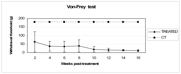
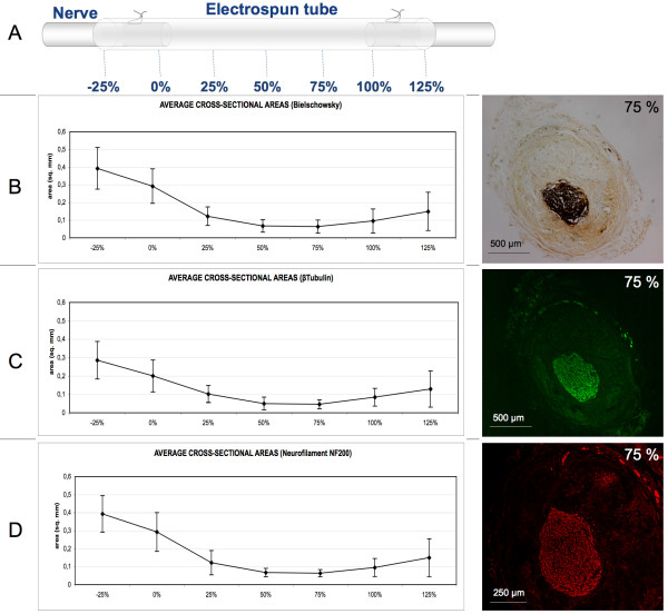
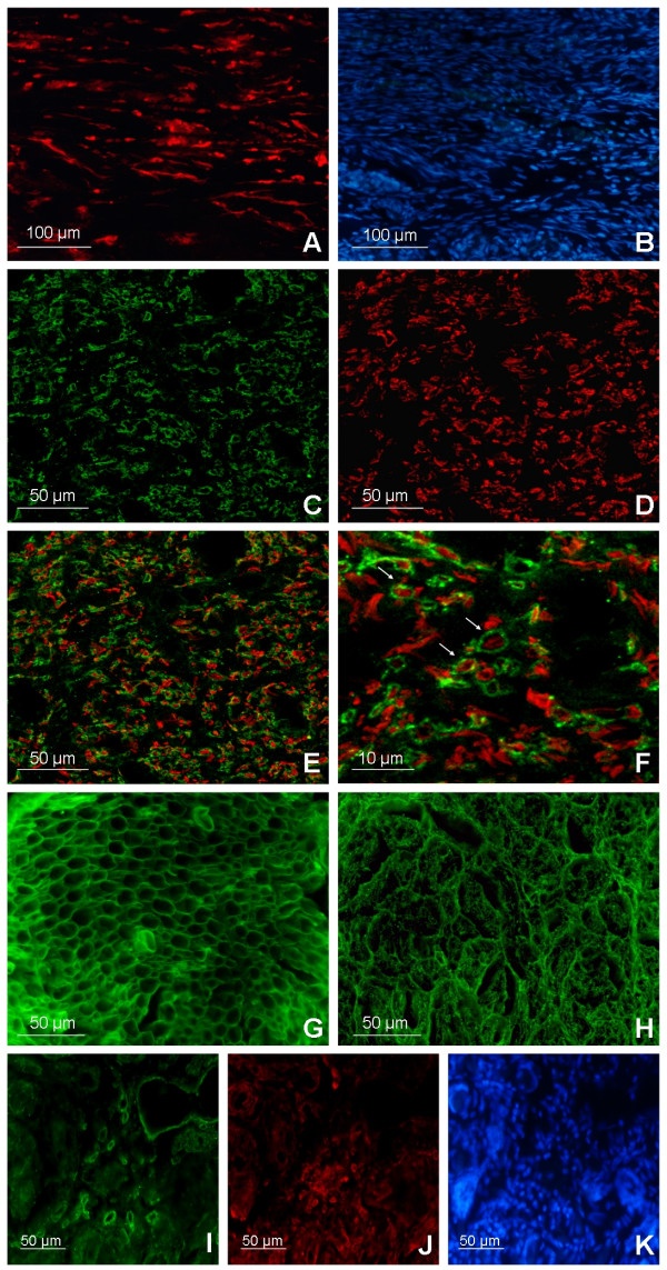
In this study we used electrospun tubes made of biodegradable polymers (a blend of PLGA/PCL) to regenerate a 10-mm nerve gap in a rat sciatic nerve in vivo. Experimental groups comprise lesioned animals (control group) and lesioned animals subjected to guide conduits implantated at the severed nerve stumps, where the tubular scaffolds are filled with saline solution. Four months after surgery, sciatic nerves failed to reconnect the two stumps of transected nerves in the control animal group. In most of the treated animals the electrospun tubes induced nervous regeneration and functional reconnection of the two severed sciatic nerve tracts. Myelination and collagen IV deposition have been detected in concurrence with regenerated fibers. No significant inflammatory response has been found. Neural tracers revealed the re-establishment of functional neuronal connections and evoked potential results showed the reinnervation of the target muscles in the majority of the treated animals.
Corroborating previous works, this study indicates that electrospun tubes, with no additional biological coating or drug loading treatment, are promising scaffolds for functional nervous regeneration. They can be knitted in meshes and various frames depending on the cytoarchitecture of the tissue to be regenerated. The versatility of this technique gives room for further scaffold improvements, like tuning the mechanical properties of the tubular structure or providing biomimetic functionalization. Moreover, these guidance conduits can be loaded with various fillers like collagen, fibrin, or self-assembling peptide gels or loaded with neurotrophic factors and seeded with cells. Electrospun scaffolds can also be synthesized in different micro-architectures to regenerate lesions in other tissues like skin and bone.
尽管近年来已经提出了许多神经假体,但在神经组织持续丧失的情况下,周围神经损伤仍然是一种创伤性病理状况,可能通过中断患者的运动感觉通路而损害其运动能力。在过去几十年中,组织工程为新方法打开了大门;然而,它们中的大多数使用刚性通道引导物,由于在患者运动期间缺乏对神经组织施加的生理局部应力,可能导致细胞损失。静电纺丝技术使得能够纺制由多种天然和合成成分组成的微纤维和纳米纤维柔性管状支架,其具有高孔隙率和显著的表面/体积比。
在本研究中,我们使用由可生物降解聚合物(PLGA/PCL混合物)制成的静电纺丝管在大鼠坐骨神经体内再生10毫米的神经间隙。实验组包括损伤动物(对照组)和在切断的神经残端植入引导导管的损伤动物,其中管状支架填充有盐水溶液。手术后四个月,对照组动物的坐骨神经未能重新连接横断神经的两个残端。在大多数接受治疗的动物中,静电纺丝管诱导了两条切断的坐骨神经束的神经再生和功能重新连接。在再生纤维同时检测到髓鞘形成和IV型胶原沉积。未发现明显的炎症反应。神经示踪剂显示功能性神经元连接的重建,诱发电位结果表明大多数接受治疗的动物中靶肌肉的再支配。
本研究证实了先前的工作,表明未经额外生物涂层或药物负载处理的静电纺丝管是用于功能性神经再生的有前景的支架。它们可以根据待再生组织的细胞结构编织成网和各种框架。该技术的多功能性为进一步改进支架提供了空间,例如调整管状结构的机械性能或提供仿生功能化。此外,这些引导导管可以装载各种填充物,如胶原蛋白、纤维蛋白或自组装肽凝胶,或装载神经营养因子并接种细胞。静电纺丝支架也可以合成不同的微结构以再生皮肤和骨骼等其他组织中的损伤。