Granot Yair, Ivorra Antoni, Rubinsky Boris
Biophysics Graduate Group, Department of Bioengineering, University of California, Berkeley, California, United States of America.
PLoS One. 2008 Apr 30;3(4):e2075. doi: 10.1371/journal.pone.0002075.
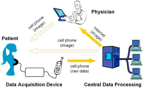
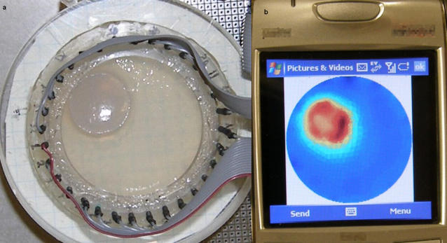
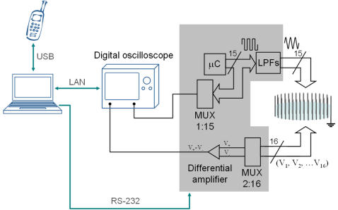
According to World Health Organization reports, some three quarters of the world population does not have access to medical imaging. In addition, in developing countries over 50% of medical equipment that is available is not being used because it is too sophisticated or in disrepair or because the health personnel are not trained to use it. The goal of this study is to introduce and demonstrate the feasibility of a new concept in medical imaging that is centered on cellular phone technology and which may provide a solution to medical imaging in underserved areas. The new system replaces the conventional stand-alone medical imaging device with a new medical imaging system made of two independent components connected through cellular phone technology. The independent units are: a) a data acquisition device (DAD) at a remote patient site that is simple, with limited controls and no image display capability and b) an advanced image reconstruction and hardware control multiserver unit at a central site. The cellular phone technology transmits unprocessed raw data from the patient site DAD and receives and displays the processed image from the central site. (This is different from conventional telemedicine where the image reconstruction and control is at the patient site and telecommunication is used to transmit processed images from the patient site). The primary goal of this study is to demonstrate that the cellular phone technology can function in the proposed mode. The feasibility of the concept is demonstrated using a new frequency division multiplexing electrical impedance tomography system, which we have developed for dynamic medical imaging, as the medical imaging modality. The system is used to image through a cellular phone a simulation of breast cancer tumors in a medical imaging diagnostic mode and to image minimally invasive tissue ablation with irreversible electroporation in a medical imaging interventional mode.
根据世界卫生组织的报告,世界上约四分之三的人口无法获得医学成像服务。此外,在发展中国家,超过50%的现有医疗设备未被使用,原因是设备过于复杂、失修,或者卫生人员没有接受过使用培训。本研究的目的是引入并证明一种以手机技术为核心的医学成像新概念的可行性,该概念可能为医疗服务不足地区的医学成像提供解决方案。新系统用一个由通过手机技术连接的两个独立组件组成的新医学成像系统取代了传统的独立医学成像设备。这两个独立单元分别是:a)远程患者端的数据采集设备(DAD),其结构简单,控制有限且无图像显示功能;b)中心端的先进图像重建和硬件控制多服务器单元。手机技术从患者端DAD传输未处理的原始数据,并从中心端接收和显示处理后的图像。(这与传统远程医疗不同,传统远程医疗中图像重建和控制在患者端,通过电信传输患者端处理后的图像)。本研究的主要目的是证明手机技术能够在所提出的模式下运行。使用我们为动态医学成像开发的一种新的频分复用电阻抗断层成像系统作为医学成像模态,来证明该概念的可行性。该系统在医学成像诊断模式下通过手机对乳腺癌肿瘤模拟进行成像,并在医学成像介入模式下对不可逆电穿孔微创组织消融进行成像。