Fu Yu, Han Jeong, Ishola Titilope, Scerbo Michelle, Adwanikar Hita, Ramsey Cara, Neugebauer Volker
Department of Neuroscience & Cell Biology, The University of Texas Medical Branch, Galveston, TX 77555-1069, USA.
Mol Pain. 2008 Jul 16;4:26. doi: 10.1186/1744-8069-4-26.
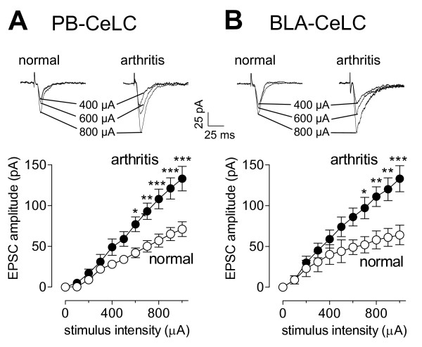
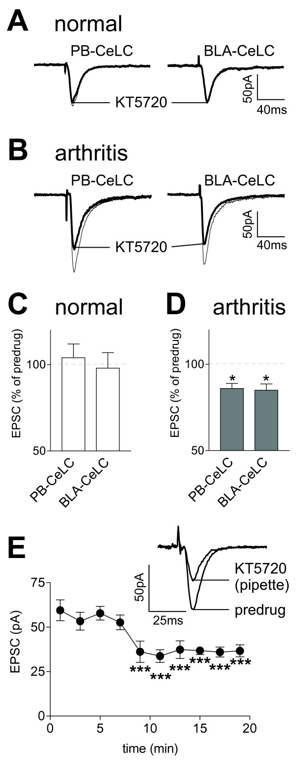
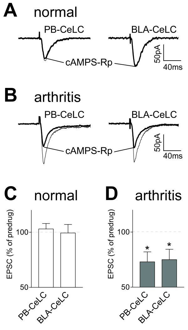
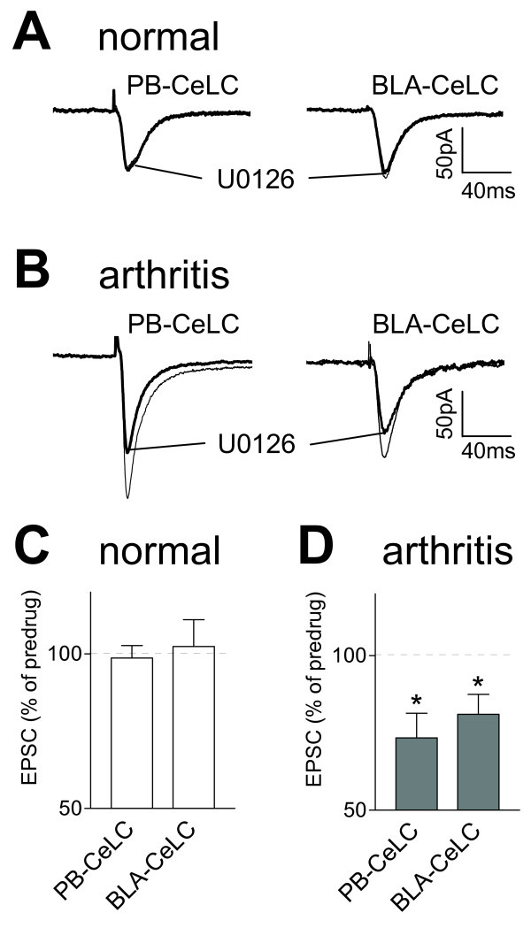
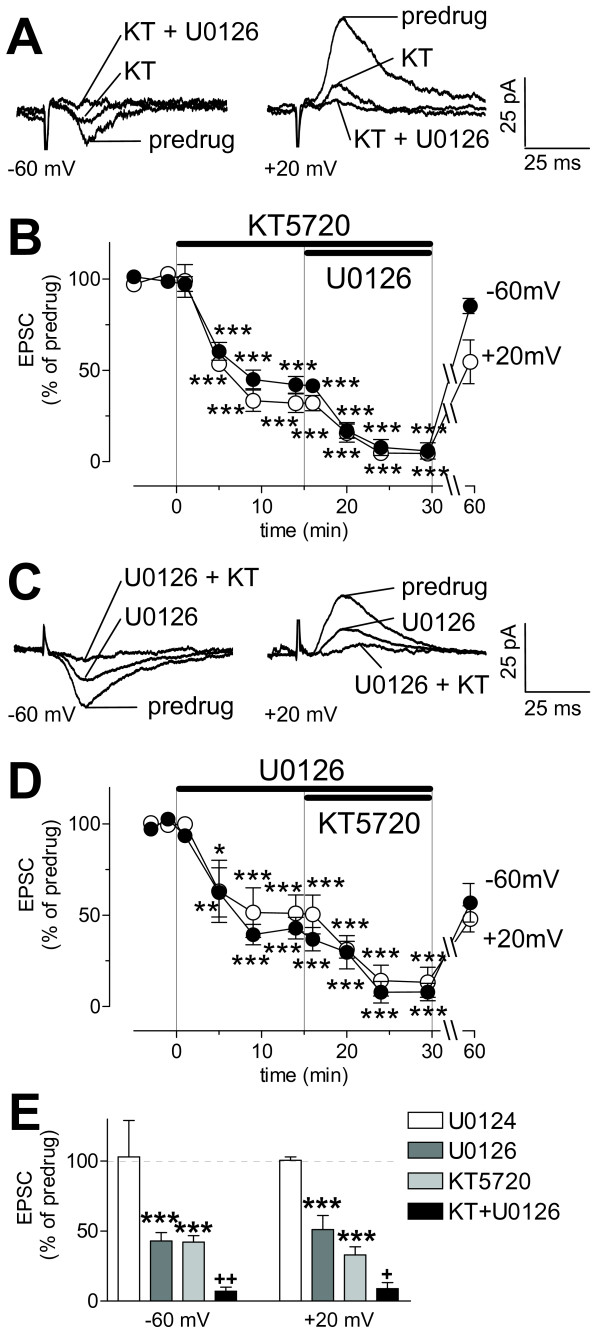
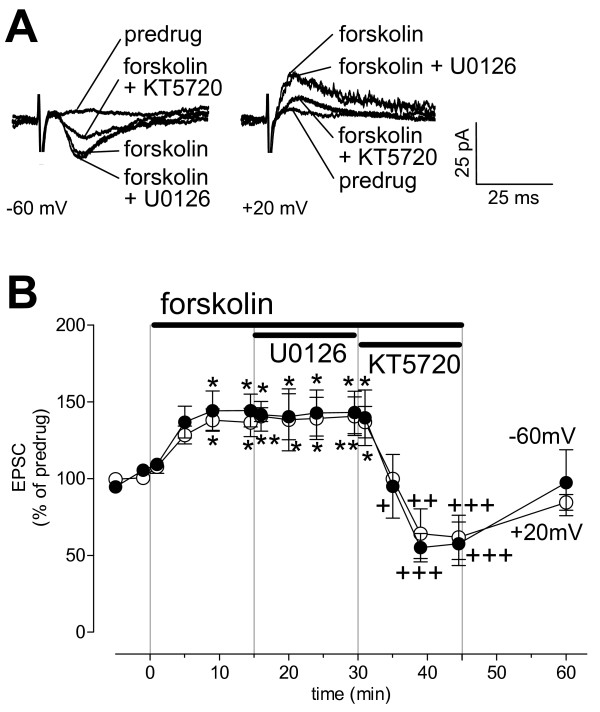
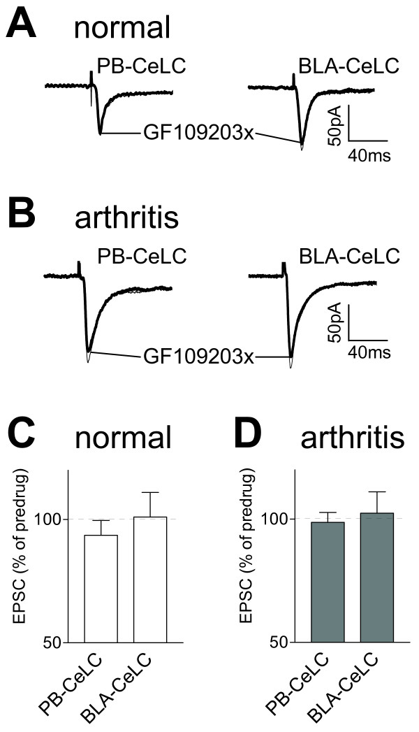
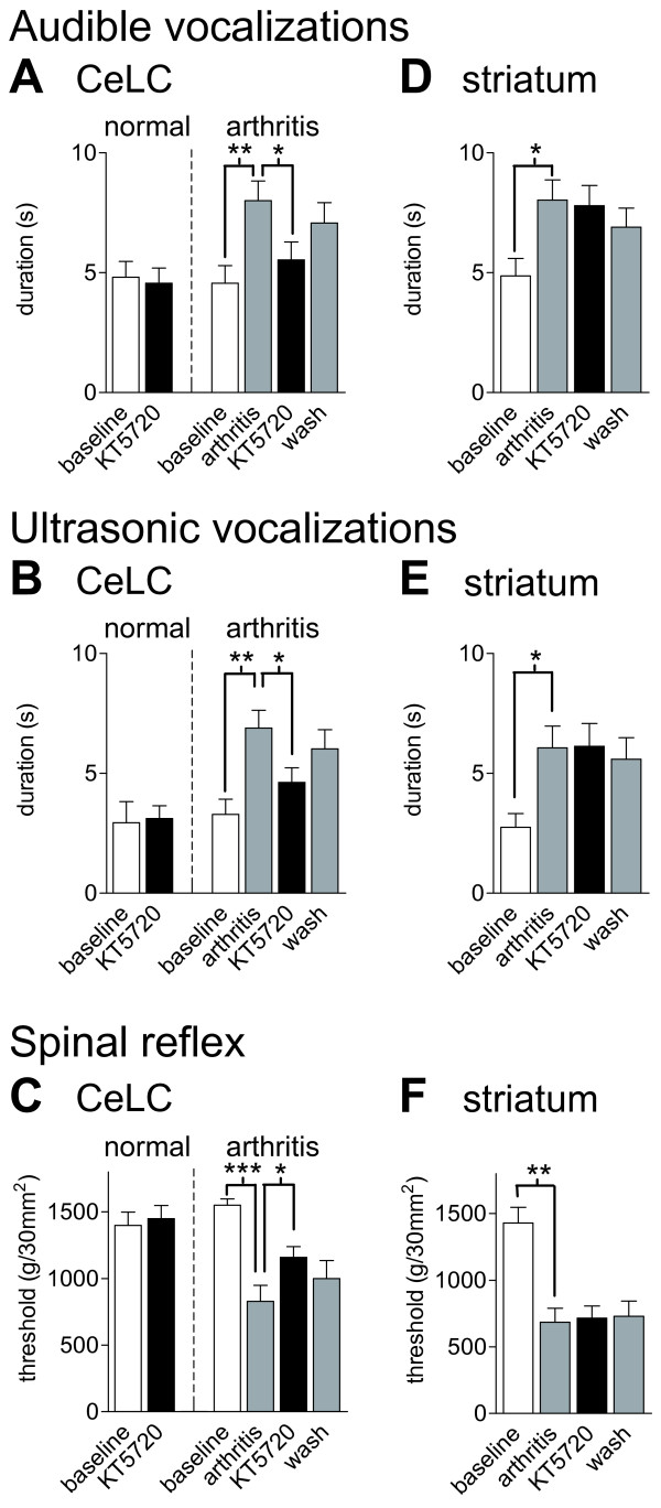
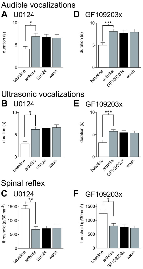
The laterocapsular division of the central nucleus of the amygdala (CeLC) has emerged as an important site of pain-related plasticity and pain modulation. Glutamate and neuropeptide receptors in the CeLC contribute to synaptic and behavioral changes in the arthritis pain model, but the intracellular signaling pathways remain to be determined. This study addressed the role of PKA, PKC, and ERK in the CeLC. Adult male Sprague-Dawley rats were used in all experiments. Whole-cell patch-clamp recordings of CeLC neurons were made in brain slices from normal rats and from rats with a kaolin/carrageenan-induced monoarthritis in the knee (6 h postinduction). Membrane-permeable inhibitors of PKA (KT5720, 1 microM; cAMPS-Rp, 10 microM) and ERK (U0126, 1 microM) activation inhibited synaptic plasticity in slices from arthritic rats but had no effect on normal transmission in control slices. A PKC inhibitor (GF109203x, 1 microM) and an inactive structural analogue of U0126 (U0124, 1 microM) had no effect. The NMDA receptor-mediated synaptic component was inhibited by KT5720 or U0126; their combined application had additive effects. U0126 did not inhibit synaptic facilitation by forskolin-induced PKA-activation. Administration of KT5720 (100 microM, concentration in microdialysis probe) or U0126 (100 microM) into the CeLC, but not striatum (placement control), inhibited audible and ultrasonic vocalizations and spinal reflexes of arthritic rats but had no effect in normal animals. GF109203x (100 microM) and U0124 (100 microM) did not affect pain behavior. The data suggest that in the amygdala PKA and ERK, but not PKC, contribute to pain-related synaptic facilitation and behavior by increasing NMDA receptor function through independent signaling pathways.
杏仁核中央核外侧囊部(CeLC)已成为疼痛相关可塑性和疼痛调节的重要部位。CeLC中的谷氨酸和神经肽受体促成了关节炎疼痛模型中的突触和行为变化,但细胞内信号通路仍有待确定。本研究探讨了蛋白激酶A(PKA)、蛋白激酶C(PKC)和细胞外信号调节激酶(ERK)在CeLC中的作用。所有实验均使用成年雄性Sprague-Dawley大鼠。在正常大鼠以及高岭土/角叉菜胶诱导的膝部单关节炎大鼠(诱导后6小时)的脑片中,对CeLC神经元进行全细胞膜片钳记录。PKA激活的膜通透性抑制剂(KT5720,1微摩尔;cAMPS-Rp,10微摩尔)和ERK激活的膜通透性抑制剂(U0126,1微摩尔)抑制了关节炎大鼠脑片的突触可塑性,但对对照脑片中的正常传递没有影响。PKC抑制剂(GF109203x,1微摩尔)和U0126的无活性结构类似物(U0124,1微摩尔)没有作用。KT5720或U0126抑制了N-甲基-D-天冬氨酸(NMDA)受体介导的突触成分;它们联合应用具有相加作用。U0126不抑制福斯高林诱导的PKA激活所引起的突触易化。将KT5720(100微摩尔,微透析探针中的浓度)或U0126(100微摩尔)注入CeLC,但不注入纹状体(位置对照),可抑制关节炎大鼠的可听和超声发声以及脊髓反射,但对正常动物没有影响。GF109203x(100微摩尔)和U0124(100微摩尔)不影响疼痛行为。数据表明,在杏仁核中,PKA和ERK而非PKC通过独立的信号通路增强NMDA受体功能,从而促成与疼痛相关的突触易化和行为。