Lawrence J R, Korber D R, Hoyle B D, Costerton J W, Caldwell D E
National Hydrology Research Institute, Environment Canada, Saskatoon, Saskatchewan.
J Bacteriol. 1991 Oct;173(20):6558-67. doi: 10.1128/jb.173.20.6558-6567.1991.
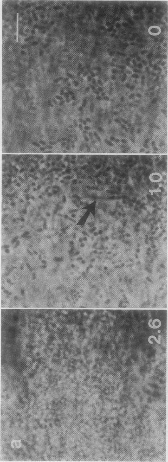
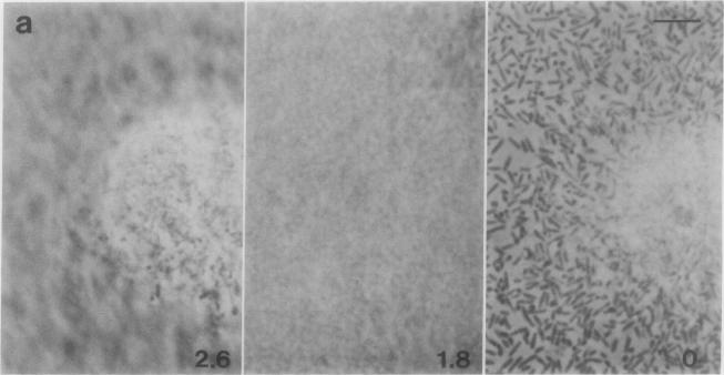
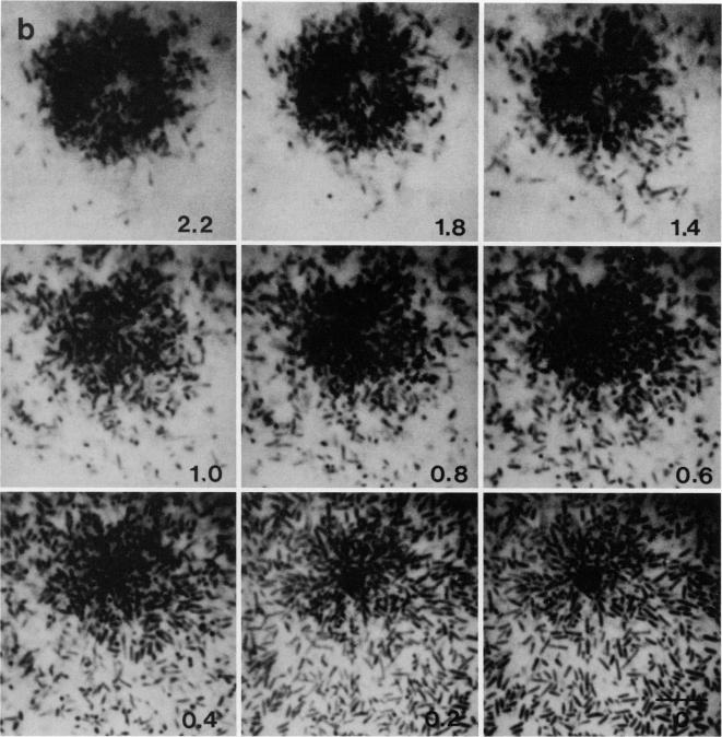
Scanning confocal laser microscopy (SCLM) was used to visualize fully hydrated microbial biofilms. The improved rejection of out-of-focus haze and the increased resolution of SCLM made it preferable to conventional phase microscopy for the analysis of living biofilms. The extent of image improvement was dependent on the characteristics of individual biofilms and was most apparent when films were dispersed in three dimensions, when they were thick, and when they contained a high number of cells. SCLM optical sections were amenable to quantitative computer-enhanced microscopy analyses, with minimal interference originating from overlying or underlying cell material. By using SCLM in conjunction with viable negative fluorescence staining techniques, horizontal (xy) and sagittal (xz) sections of intact biofilms of Pseudomonas aeruginosa, Pseudomonas fluorescens, and Vibrio parahaemolyticus were obtained. These optical sections were then analyzed by image-processing techniques to assess the distribution of cellular and noncellular areas within the biofilm matrices. The Pseudomonas biofilms were most cell dense at their attachment surfaces and became increasingly diffuse near their outer regions, whereas the Vibrio biofilms exhibited the opposite trend. Biofilms consisting of different species exhibited distinctive arrangements of the major biofilm structural components (cellular and extracellular materials and space). In general, biofilms were found to be highly hydrated, open structures composed of 73 to 98% extracellular materials and space. The use of xz sectioning revealed more detail of biofilm structure, including the presence of large void spaces within the Vibrio biofilms. In addition, three-dimensional reconstructions of biofilms were constructed and were displayed as stereo pairs. Application of the concepts of architectural analysis to mixed- or pure-species biofilms will allow detailed examination of the relationships among biofilm structure, adaptation, and response to stress.
扫描共聚焦激光显微镜(SCLM)用于观察完全水合的微生物生物膜。SCLM对离焦雾度的改善以及分辨率的提高,使其在分析活生物膜方面优于传统相差显微镜。图像改善的程度取决于单个生物膜的特性,当生物膜在三维空间中分散、较厚且含有大量细胞时最为明显。SCLM光学切片适用于定量计算机增强显微镜分析,来自上层或下层细胞物质的干扰最小。通过将SCLM与活菌负荧光染色技术结合使用,获得了铜绿假单胞菌、荧光假单胞菌和副溶血性弧菌完整生物膜的水平(xy)和矢状(xz)切片。然后通过图像处理技术对这些光学切片进行分析,以评估生物膜基质中细胞和非细胞区域的分布。假单胞菌生物膜在其附着表面细胞密度最高,在其外部区域附近变得越来越分散,而弧菌生物膜则呈现相反的趋势。由不同物种组成的生物膜表现出主要生物膜结构成分(细胞和细胞外物质及空间)的独特排列。一般来说,生物膜被发现是高度水合的开放结构,由73%至98%的细胞外物质和空间组成。使用xz切片揭示了生物膜结构的更多细节,包括弧菌生物膜中存在的大空隙。此外,构建了生物膜的三维重建并以立体对的形式显示。将结构分析的概念应用于混合或纯物种生物膜将允许详细检查生物膜结构、适应性和对压力的反应之间的关系。