Zhu Wei-Zhong, Santana Luis F, Laflamme Michael A
Department of Pathology, University of Washington, Seattle, Washington, United States of America.
PLoS One. 2009;4(4):e5407. doi: 10.1371/journal.pone.0005407. Epub 2009 Apr 30.
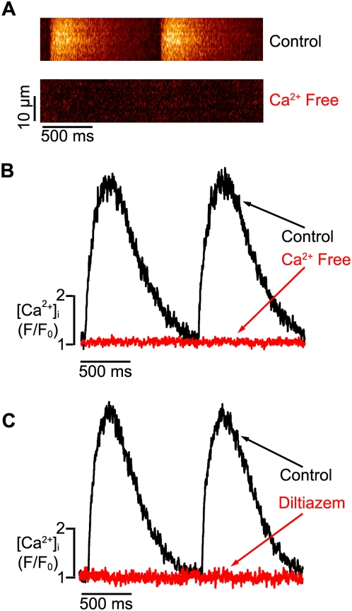
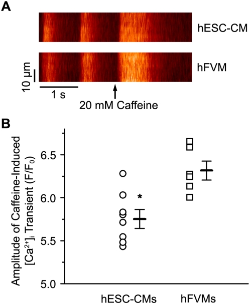
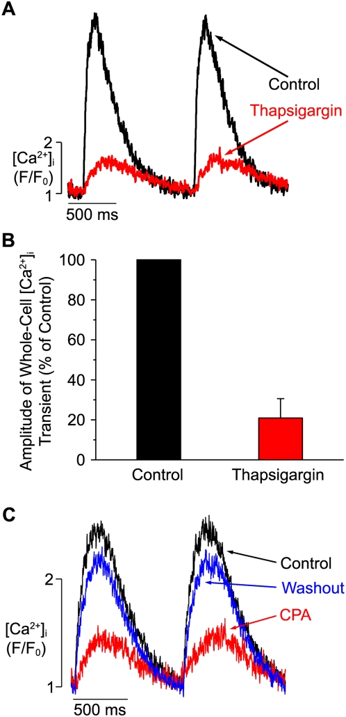
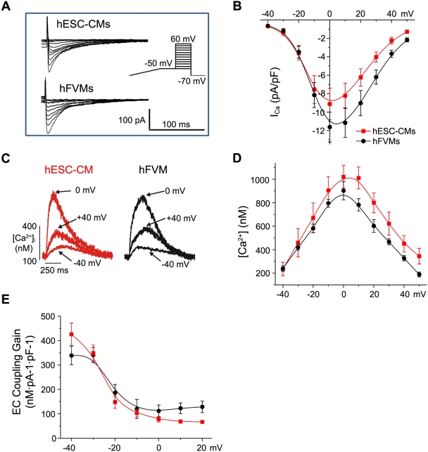
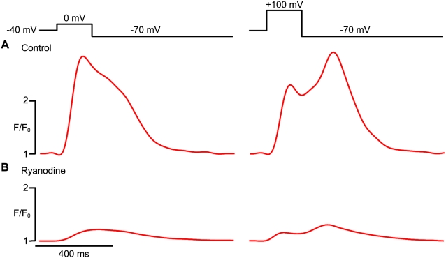
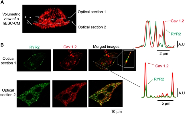
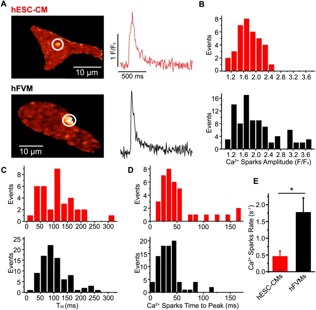
We investigated the mechanisms of excitation-contraction (EC) coupling in human embryonic stem cell-derived cardiomyocytes (hESC-CMs) and fetal ventricular myocytes (hFVMs) using patch-clamp electrophysiology and confocal microscopy. We tested the hypothesis that Ca(2+) influx via voltage-gated L-type Ca(2+) channels activates Ca(2+) release from the sarcoplasmic reticulum (SR) via a local control mechanism in hESC-CMs and hFVMs. Field-stimulated, whole-cell Ca(2+) transients in hESC-CMs required Ca(2+) entry through L-type Ca(2+) channels, as evidenced by the elimination of such transients by either removal of extracellular Ca(2+) or treatment with diltiazem, an L-type channel inhibitor. Ca(2+) release from the SR also contributes to the Ca(2+) transient in these cells, as evidenced by studies with drugs interfering with either SR Ca(2+) release (i.e. ryanodine and caffeine) or reuptake (i.e. thapsigargin and cyclopiazonic acid). As in adult ventricular myocytes, membrane depolarization evoked large L-type Ca(2+) currents (I(Ca)) and corresponding whole-cell Ca(2+) transients in hESC-CMs and hFVMs, and the amplitude of both I(Ca) and the Ca(2+) transients were finely graded by the magnitude of the depolarization. hESC-CMs exhibit a decreasing EC coupling gain with depolarization to more positive test potentials, "tail" Ca(2+) transients upon repolarization from extremely positive test potentials, and co-localized ryanodine and sarcolemmal L-type Ca(2+) channels, all findings that are consistent with the local control hypothesis. Finally, we recorded Ca(2+) sparks in hESC-CMs and hFVMs. Collectively, these data support a model in which tight, local control of SR Ca(2+) release by the I(Ca) during EC coupling develops early in human cardiomyocytes.
我们使用膜片钳电生理学和共聚焦显微镜研究了人胚胎干细胞衍生的心肌细胞(hESC-CMs)和胎儿心室肌细胞(hFVMs)中兴奋-收缩(EC)偶联的机制。我们检验了以下假设:通过电压门控L型钙通道的Ca(2+)内流通过局部控制机制激活hESC-CMs和hFVMs中肌浆网(SR)的Ca(2+)释放。hESC-CMs中的场刺激全细胞Ca(2+)瞬变需要Ca(2+)通过L型钙通道进入,去除细胞外Ca(2+)或用L型通道抑制剂地尔硫卓处理消除此类瞬变即可证明这一点。SR的Ca(2+)释放也有助于这些细胞中的Ca(2+)瞬变,干扰SR Ca(2+)释放(即ryanodine和咖啡因)或再摄取(即毒胡萝卜素和平圆二烯酸)的药物研究证明了这一点。与成人心室肌细胞一样,膜去极化在hESC-CMs和hFVMs中诱发了大的L型钙电流(I(Ca))和相应的全细胞Ca(2+)瞬变,I(Ca)和Ca(2+)瞬变的幅度均由去极化幅度精细分级。hESC-CMs在去极化到更正的测试电位时表现出EC偶联增益降低,从极正的测试电位复极化时出现“尾”Ca(2+)瞬变,以及ryanodine和肌膜L型钙通道共定位,所有这些发现都与局部控制假设一致。最后,我们记录了hESC-CMs和hFVMs中的Ca(2+)火花。总的来说,这些数据支持了一个模型,即在EC偶联过程中,I(Ca)对SR Ca(2+)释放的紧密局部控制在人类心肌细胞早期就已形成。