Bastian Susanne, Busch Wibke, Kühnel Dana, Springer Armin, Meissner Tobias, Holke Roland, Scholz Stefan, Iwe Maria, Pompe Wolfgang, Gelinsky Michael, Potthoff Annegret, Richter Volkmar, Ikonomidou Chrysanthy, Schirmer Kristin
Department of Pediatric Neurology, University Children's Hospital Carl Gustav Carus, University of Technology Dresden, Dresden, Germany.
Environ Health Perspect. 2009 Apr;117(4):530-6. doi: 10.1289/ehp.0800121. Epub 2008 Dec 1.
Tungsten carbide nanoparticles are being explored for their use in the manufacture of hard metals. To develop nanoparticles for broad applications, potential risks to human health and the environment should be evaluated and taken into consideration.
We aimed to assess the toxicity of well-characterized tungsten carbide (WC) and cobalt-doped tungsten carbide (WC-Co) nanoparticle suspensions in an array of mammalian cells.
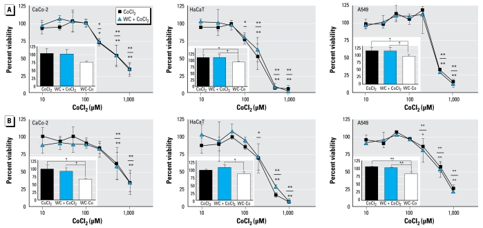
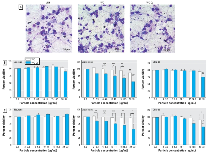
We examined acute toxicity of WC and of WC-Co (10% weight content Co) nanoparticles in different human cell lines (lung, skin, and colon) as well as in rat neuronal and glial cells (i.e., primary neuronal and astroglial cultures and the oligodendrocyte precursor cell line OLN-93). Furthermore, using electron microscopy, we assessed whether nanoparticles can be taken up by living cells. We chose these in vitro systems in order to evaluate for potential toxicity of the nanoparticles in different mammalian organs (i.e., lung, skin, intestine, and brain).
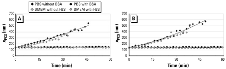
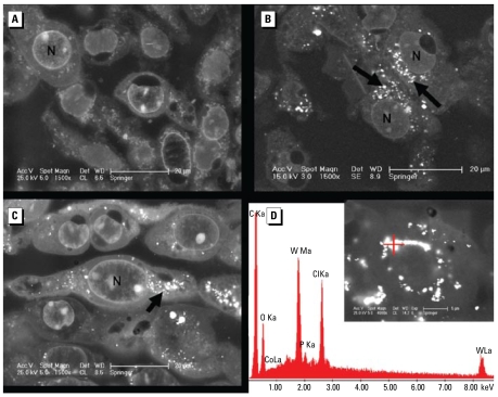
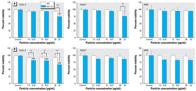
Chemical-physical characterization confirmed that WC as well as WC-Co nanoparticles with a mean particle size of 145 nm form stable suspensions in serum-containing cell culture media. WC nanoparticles were not acutely toxic to the studied cell lines. However, cytotoxicity became apparent when particles were doped with Co. The most sensitive were astrocytes and colon epithelial cells. Cytotoxicity of WC-Co nanoparticles was higher than expected based on the ionic Co content of the particles. Analysis by electron microscopy demonstrated presence of WC nanoparticles within mammalian cells.
Our findings demonstrate that doping of WC nanoparticles with Co markedly increases their cytotoxic effect and that the presence of WC-Co in particulate form is essential to elicit this combinatorial effect.
碳化钨纳米颗粒正被探索用于硬质金属制造。为开发可广泛应用的纳米颗粒,应评估并考虑其对人类健康和环境的潜在风险。
我们旨在评估特征明确的碳化钨(WC)和钴掺杂碳化钨(WC-Co)纳米颗粒悬浮液对一系列哺乳动物细胞的毒性。
我们检测了WC和WC-Co(钴重量含量为10%)纳米颗粒对不同人类细胞系(肺、皮肤和结肠)以及大鼠神经元和神经胶质细胞(即原代神经元和星形胶质细胞培养物以及少突胶质前体细胞系OLN-93)的急性毒性。此外,我们使用电子显微镜评估纳米颗粒是否能被活细胞摄取。我们选择这些体外系统以评估纳米颗粒在不同哺乳动物器官(即肺、皮肤、肠道和脑)中的潜在毒性。
化学物理特性证实,平均粒径为145 nm的WC以及WC-Co纳米颗粒在含血清的细胞培养基中形成稳定悬浮液。WC纳米颗粒对所研究的细胞系无急性毒性。然而,当颗粒掺杂钴时,细胞毒性变得明显。最敏感的是星形胶质细胞和结肠上皮细胞。WC-Co纳米颗粒的细胞毒性高于基于颗粒离子钴含量的预期。电子显微镜分析表明哺乳动物细胞内存在WC纳米颗粒。
我们的数据表明,WC纳米颗粒掺杂钴会显著增加其细胞毒性作用,且颗粒形式的WC-Co的存在对于引发这种联合效应至关重要。