Yousef Zaheer R, Foley Paul W X, Khadjooi Kayvan, Chalil Shajil, Sandman Harald, Mohammed Noor U H, Leyva Francisco
Department of Cardiology, Good Hope Hospital, Sutton Coldfield, West Midlands, UK.
BMC Cardiovasc Disord. 2009 Aug 9;9:37. doi: 10.1186/1471-2261-9-37.
It is apparent that despite lack of family history, patients with the morphological characteristics of left ventricular non-compaction develop arrhythmias, thrombo-embolism and left ventricular dysfunction.
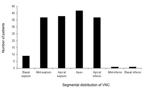
Forty two patients, aged 48.7 +/- 2.3 yrs (mean +/- SEM) underwent cardiovascular magnetic resonance (CMR) for the quantification of left ventricular volumes and extent of non-compacted (NC) myocardium. The latter was quantified using planimetry on the two-chamber long axis LV view (NC area). The patients included those referred specifically for CMR to investigate suspected cardiomyopathy, and as such is represents a selected group of patients.
At presentation, 50% had dyspnoea, 19% chest pain, 14% palpitations and 5% stroke. Pulmonary embolism had occurred in 7% and brachial artery embolism in 2%. The ECG was abnormal in 81% and atrial fibrillation occurred in 29%. Transthoracic echocardiograms showed features of NC in only 10%. On CMR, patients who presented with dyspnoea had greater left ventricular volumes (both p < 0.0001) and a lower left ventricular ejection fraction (LVEF) (p < 0.0001) than age-matched, healthy controls. In patients without dyspnoea (n = 21), NC area correlated positively with end-diastolic volume (r = 0.52, p = 0.0184) and end-systolic volume (r = 0.56, p = 0.0095), and negatively with EF (r = -0.72, p = 0.0001).
Left ventricular non-compaction is associated with dysrrhythmias, thromboembolic events, chest pain and LV dysfunction. The inverse correlation between NC area and EF suggests that NC contributes to left ventricular dysfunction.
显然,尽管缺乏家族病史,但具有左心室心肌致密化不全形态学特征的患者仍会发生心律失常、血栓栓塞和左心室功能障碍。
42名年龄为48.7±2.3岁(平均±标准误)的患者接受了心血管磁共振成像(CMR)检查,以量化左心室容积和心肌致密化不全(NC)的范围。后者通过在左心室两腔长轴视图上进行面积测量法(NC面积)来量化。这些患者包括专门因怀疑患有心肌病而转诊进行CMR检查的患者,因此这代表了一组经过挑选的患者。
就诊时,50%的患者有呼吸困难,19%有胸痛,14%有心悸,5%有中风。7%的患者发生过肺栓塞,2%的患者发生过肱动脉栓塞。81%的患者心电图异常,29%的患者发生心房颤动。经胸超声心动图仅显示10%的患者有NC特征。在CMR检查中,出现呼吸困难的患者与年龄匹配的健康对照组相比,左心室容积更大(均p<0.0001),左心室射血分数(LVEF)更低(p<0.0001)。在没有呼吸困难的患者(n=21)中,NC面积与舒张末期容积呈正相关(r=0.52,p=0.0184)和收缩末期容积呈正相关(r=0.56,p=0.0095),与EF呈负相关(r=-0.72,p=0.0001)。
左心室心肌致密化不全与心律失常、血栓栓塞事件、胸痛和左心室功能障碍有关。NC面积与EF之间的负相关表明NC导致左心室功能障碍。