He Jing, Farias Sarah, Martinez Oliver, Reed Bruce, Mungas Dan, Decarli Charles
Department of Neurology, and the Imaging of Dementia and Aging Laboratory, Center for Neuroscience Preventive Medicine, University of California at Davis, Sacramento, CA 95817, USA.
Arch Neurol. 2009 Nov;66(11):1393-9. doi: 10.1001/archneurol.2009.252.
To evaluate demographics, magnetic resonance imaging (MRI) measures, and vascular risk among mild cognitive impairment (MCI) subtypes.
Cross-sectional study.
Both clinics and the community.
A total of 153 subjects with MCI, 218 cognitively normal older individuals (controls), and 68 patients with Alzheimer disease.
Classification of subjects with MCI according to current subtype diagnostic convention based on neuropsychological performance, estimates of vascular risk based on medical history, research MRI unless there was a specific contraindication, and apolipoprotein E genotype.
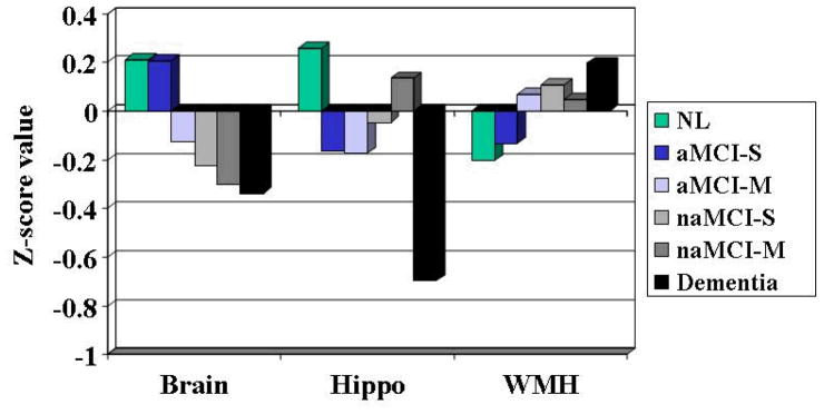
Of the 153 subjects with MCI, 65 were diagnosed with amnestic single-domain, 46 with amnestic multiple-domain, 27 with nonamnestic single-domain, and 15 with nonamnestic multiple-domain MCI. Analyses of control, MCI, and Alzheimer disease cases revealed significant differences in brain and hippocampal volumes between each group. Post hoc analyses of MRI measures among the MCI subtypes found that patients with amnestic single-domain MCI had significantly less brain atrophy and that hippocampal volume differed significantly from controls for the 2 amnestic forms of MCI. Apolipoprotein E genotype prevalence was significantly greater in the amnestic and nonamnestic subtypes of MCI. Conversely, the nonamnestic subtypes were more likely to have increased vascular risk and to be African American.
Amnestic forms of MCI appear to have demographic, genetic, and MRI findings suggestive of Alzheimer disease pathology, whereas the nonamnestic forms of MCI have findings suggestive of vascular disease. Importantly, however, all subjects with MCI showed evidence of brain injury, and the biological differences among subtypes are relatively subtle beyond the memory vs nonmemory groupings.
评估轻度认知障碍(MCI)各亚型的人口统计学特征、磁共振成像(MRI)测量指标及血管风险。
横断面研究。
诊所及社区。
共153例MCI受试者、218例认知正常的老年人(对照组)以及68例阿尔茨海默病患者。
根据基于神经心理学表现的当前亚型诊断标准对MCI受试者进行分类,根据病史评估血管风险,除非有特定禁忌证,否则进行研究性MRI检查,以及载脂蛋白E基因型。
在153例MCI受试者中,65例被诊断为遗忘型单领域MCI,46例为遗忘型多领域MCI,27例为非遗忘型单领域MCI,15例为非遗忘型多领域MCI。对对照组、MCI组和阿尔茨海默病病例的分析显示,各组之间脑体积和海马体积存在显著差异。对MCI各亚型MRI测量指标的事后分析发现,遗忘型单领域MCI患者的脑萎缩明显较少,且两种遗忘型MCI的海马体积与对照组有显著差异。载脂蛋白E基因型在MCI的遗忘型和非遗忘型亚型中的患病率显著更高。相反,非遗忘型亚型更有可能有血管风险增加且为非裔美国人。
遗忘型MCI似乎在人口统计学、遗传学和MRI表现上提示有阿尔茨海默病病理特征,而非遗忘型MCI的表现提示有血管疾病。然而,重要的是,所有MCI受试者均显示有脑损伤证据,且各亚型之间的生物学差异在记忆组与非记忆组分类之外相对细微。