Page Thomas D P, King B, Stephens T, Dingle J T
Strangeways Research Laboratory, Cambridge, UK.
Ann Rheum Dis. 1991 Feb;50(2):75-80. doi: 10.1136/ard.50.2.75.
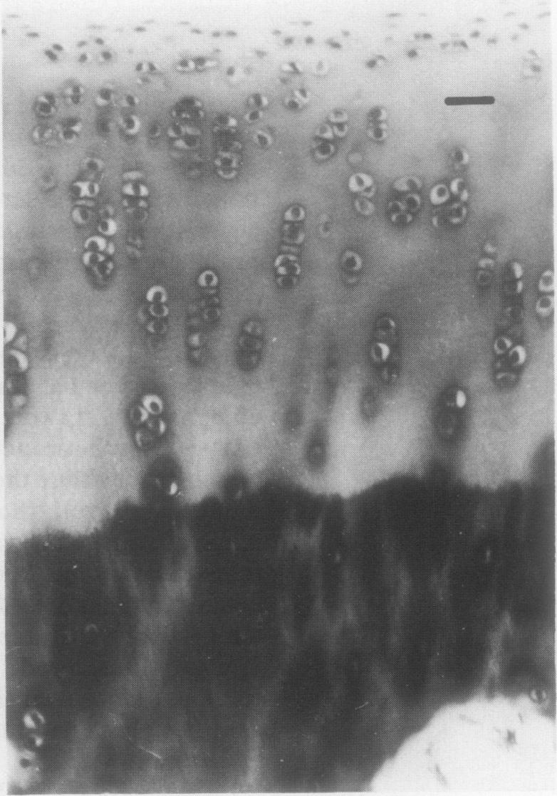
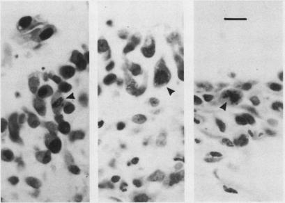
The response of the rabbit knee joint to a brief episode of cytokine induced damage is described. After three intra-articular injections of catabolin/interleukin-1 all joint cartilages showed an immediate extensive loss of proteoglycan (glycosaminoglycan), which was gradually replaced over three to four weeks. Glycosaminoglycan biosynthesis (measured by 35SO4 uptake) was initially depressed, but at one week had almost doubled its rate as compared with the normal side. This increased synthetic activity was further maintained throughout the duration of the experiment (28 days), though the rate gradually fell. Histological cartilage metachromasia to toluidine blue mirrored the glycosaminoglycan changes. No disturbance of the articular cartilage collagen network was found. It is considered, therefore, that during treatment for arthritis the indigenous chondrocyte must continue to be capable of carrying out regenerative matrix repair and that antiarthritic agents should first be screened for interference with that process.
本文描述了兔膝关节对细胞因子诱导的短暂损伤的反应。在关节内注射三次分解代谢素/白细胞介素-1后,所有关节软骨均立即出现蛋白聚糖(糖胺聚糖)大量流失,这种流失在三到四周内逐渐得到补充。糖胺聚糖生物合成(通过35SO4摄取量测定)最初受到抑制,但在一周时与正常侧相比其速率几乎增加了一倍。尽管速率逐渐下降,但在整个实验期间(28天)这种增加的合成活性得以进一步维持。组织学上软骨对甲苯胺蓝的异染性反映了糖胺聚糖的变化。未发现关节软骨胶原网络受到干扰。因此,可以认为在关节炎治疗期间,内源性软骨细胞必须继续具备进行再生性基质修复的能力,并且抗关节炎药物应首先进行筛选,以确定其是否会干扰该过程。