Department of Microbiology and Immunology, Louisiana State University Health Sciences Center - Shreveport, 1501 Kings Highway, Shreveport, LA 71130, USA.
BMC Microbiol. 2009 Dec 14;9:258. doi: 10.1186/1471-2180-9-258.
Helicobacter pylori specifically takes up cholesterol and incorporates it into the bacterial membrane, yet little is currently known about cholesterol's physiological roles. We compared phenotypes and in vivo colonization ability of H. pylori grown in a defined, serum-free growth medium, F12 with 1 mg/ml albumin containing 0 to 50 mug/ml cholesterol.
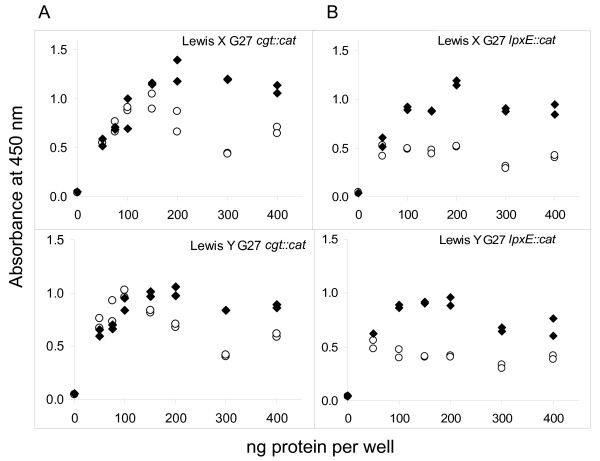
While doubling times were largely unaffected by cholesterol, other overt phenotypic changes were observed. H. pylori strain SS1 grown in defined medium with cholesterol successfully colonized the stomach of gerbils, whereas SS1 grown without cholesterol failed to colonize. H. pylori lipopolysaccharide often displays Lewis X and/or Y antigens. Expression of these antigens measured by whole-cell ELISA was markedly enhanced in response to growth of strain SS1, 26695, or G27 in cholesterol. In addition, electrophoretic analysis of lipopolysaccharide in wild type G27 and in mutants lacking the O-chain revealed structural changes within the oligosaccharide core/lipid A moieties. These responses in Lewis antigen levels and in lipopolysaccharide profiles to cholesterol availability were highly specific, because no changes took place when cholesterol was substituted by beta-sitosterol or bile salts. Disruption of the genes encoding cholesterol alpha-glucosyltransferase or lipid A phosphoethanolamine transferase had no effect on Lewis expression, nor on lipopolysaccharide profiles, nor on the cholesterol responsiveness of these properties. Disruption of the lipid A 1-phosphatase gene eliminated the effect of cholesterol on lipopolysaccharide profiles but not its effect on Lewis expression.
Together these results suggest that cholesterol depletion leads to aberrant forms of LPS that are dependent upon dephosphorylation of lipid A at the 1-position. A tentative model for the observed effects of cholesterol is discussed in which sequential steps of lipopolysaccharide biogenesis and, independently, presentation of Lewis antigen at the cell surface, depend upon membrane composition. These new findings demonstrate that cholesterol availability permits H. pylori to modify its cell envelope in ways that can impact colonization of host tissue in vivo.
幽门螺杆菌专门摄取胆固醇并将其纳入细菌膜,但目前对胆固醇的生理作用知之甚少。我们比较了在无血清的 F12 生长培养基中生长的、含有 1 毫克/毫升白蛋白的幽门螺杆菌,其中胆固醇的浓度为 0 至 50 微克/毫升,其表型和体内定植能力。
虽然胆固醇对倍增时间影响不大,但观察到其他明显的表型变化。在含有胆固醇的定义培养基中生长的 SS1 菌株成功定植了沙鼠的胃,而没有胆固醇的 SS1 菌株则无法定植。幽门螺杆菌的脂多糖通常显示 Lewis X 和/或 Y 抗原。通过全细胞 ELISA 测量这些抗原的表达,在 SS1、26695 或 G27 菌株在胆固醇中的生长反应明显增强。此外,野生型 G27 的脂多糖的电泳分析以及缺乏 O-链的突变体显示,寡糖核心/脂酰 A 部分的结构发生了变化。这些对胆固醇可用性的 Lewis 抗原水平和脂多糖谱的反应是高度特异的,因为当用 β-谷甾醇或胆汁盐替代胆固醇时,没有发生变化。胆固醇 α-葡萄糖基转移酶或脂酰 A 磷酸乙醇胺转移酶基因的破坏对 Lewis 表达、脂多糖谱或这些特性对胆固醇的反应性没有影响。脂质 A 1-磷酸酶基因的破坏消除了胆固醇对脂多糖谱的影响,但没有消除其对 Lewis 表达的影响。
这些结果表明,胆固醇耗竭导致 LPS 的异常形式,这些形式依赖于脂质 A 在 1 位的去磷酸化。讨论了观察到的胆固醇效应的一个试探性模型,其中脂多糖生物发生的连续步骤,以及独立地,细胞表面上 Lewis 抗原的呈现,取决于膜组成。这些新发现表明,胆固醇的可用性使幽门螺杆菌能够改变其细胞包膜,从而影响其在体内定植宿主组织的能力。