Department of Psychology, Life Sciences Centre, Dalhousie University, Halifax, Nova Scotia, Canada.
J Comp Neurol. 2010 Apr 1;518(7):1133-55. doi: 10.1002/cne.22268.
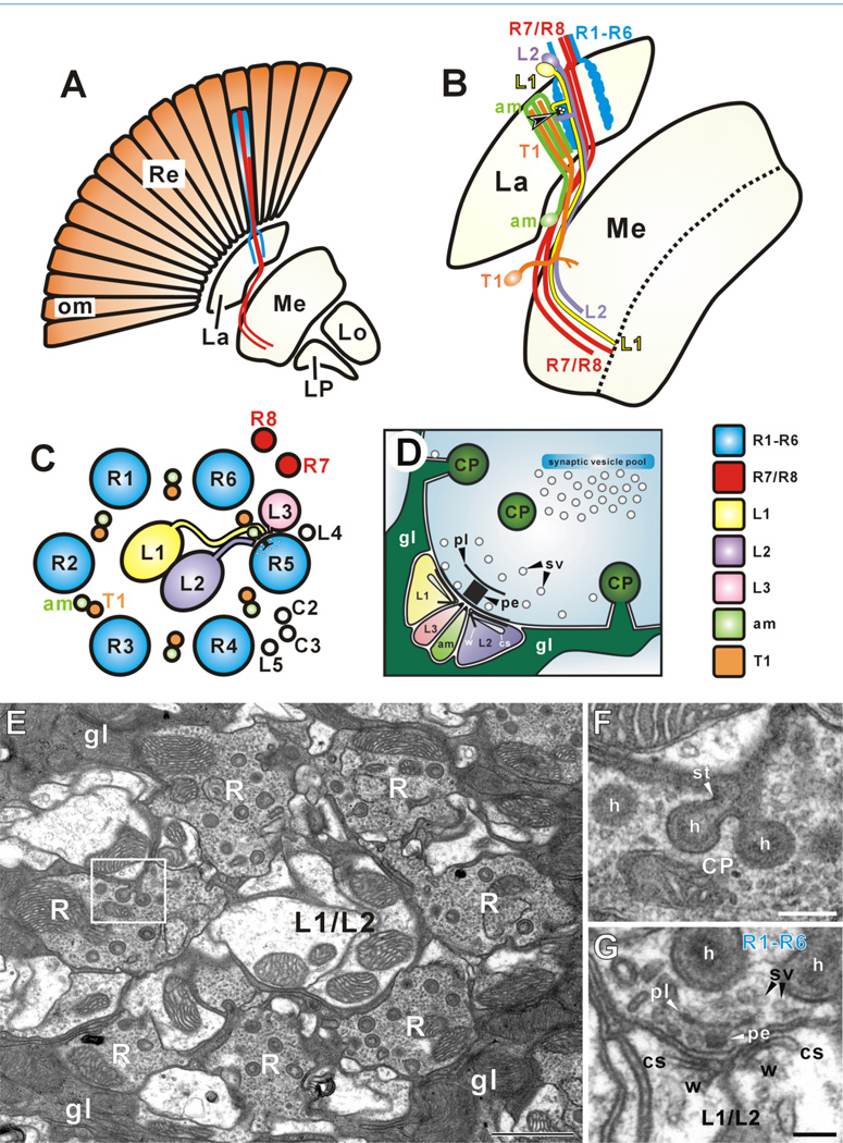
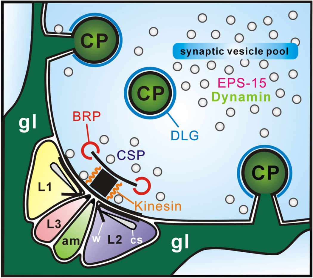
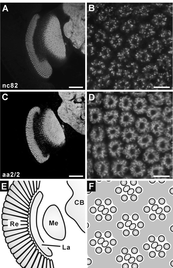
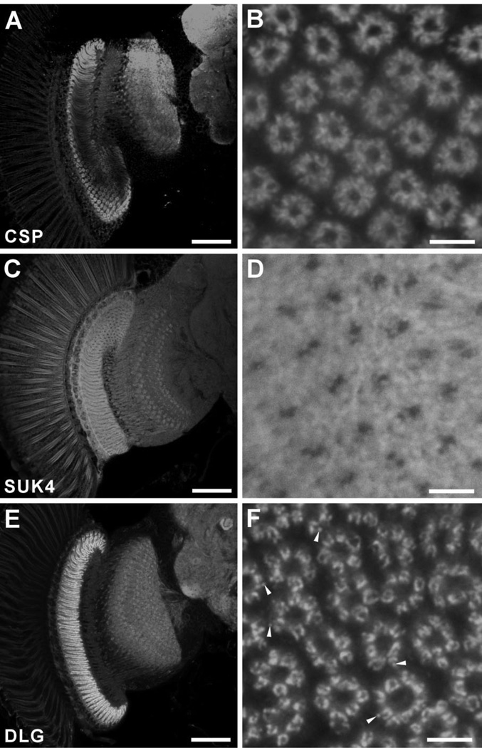
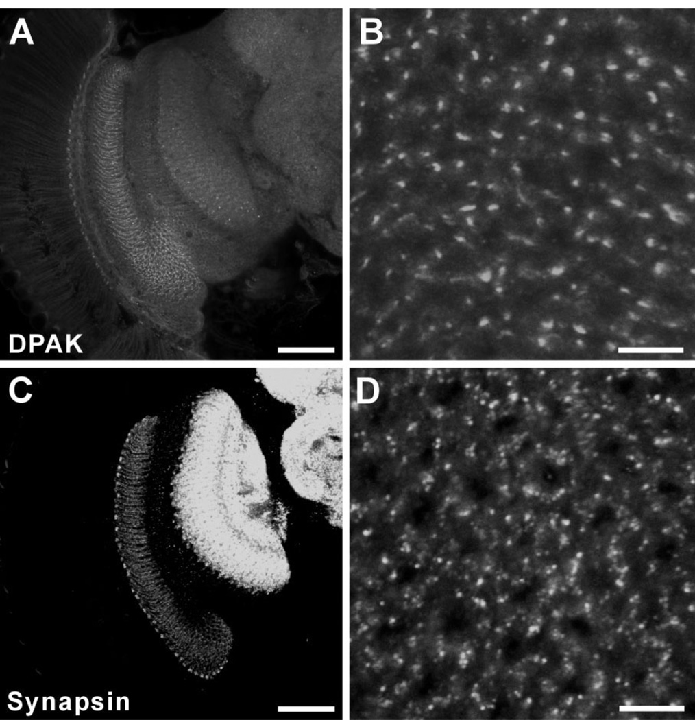
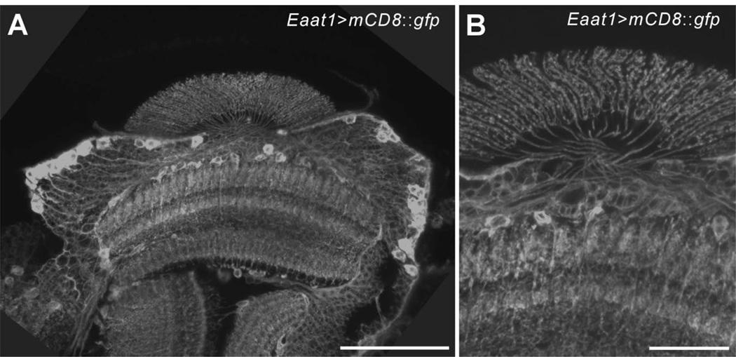
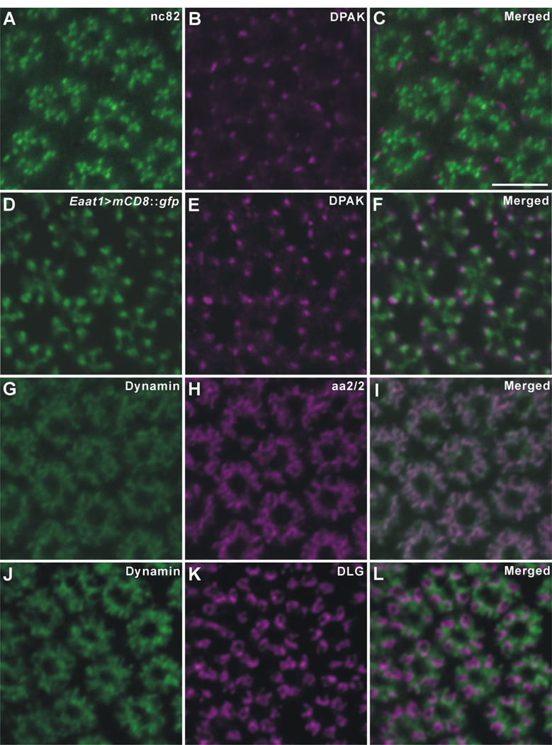
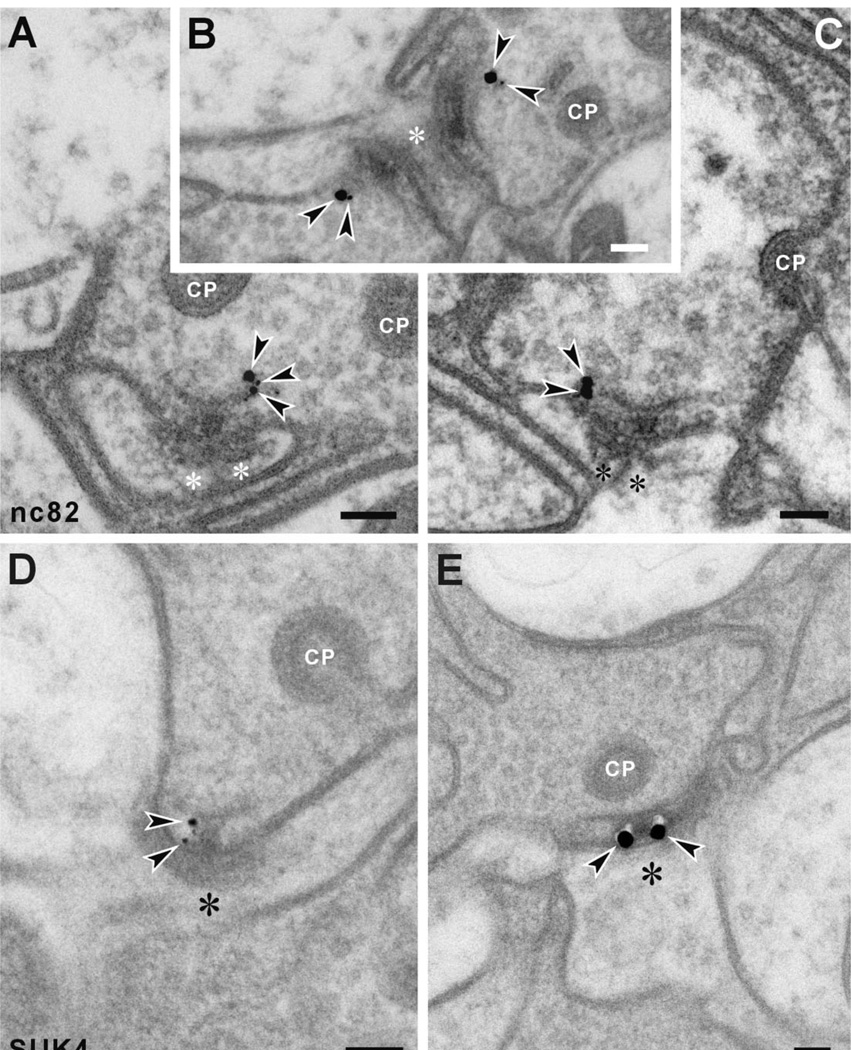
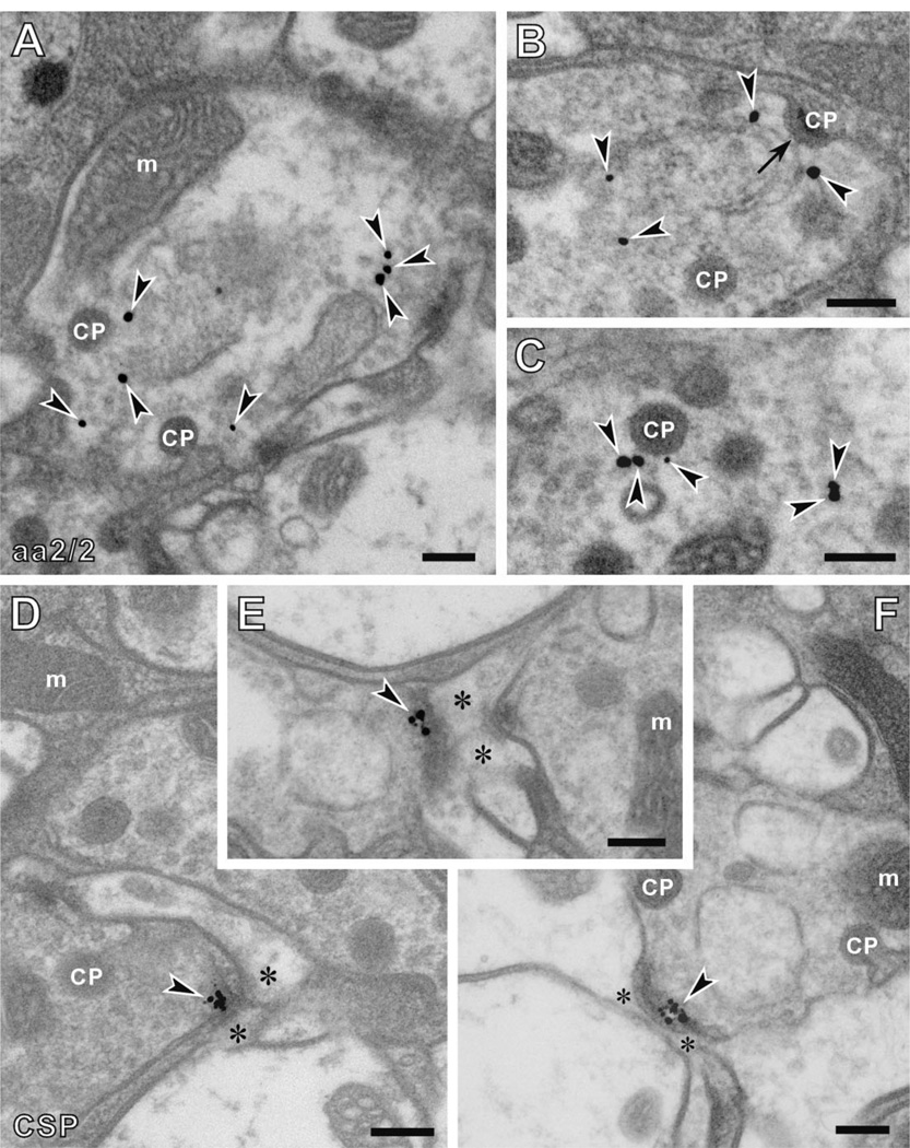
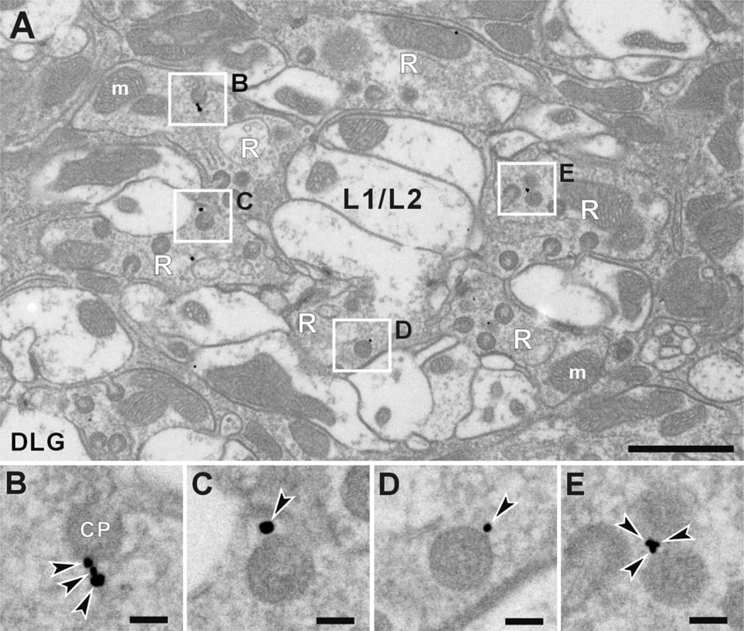
The location of proteins that contribute to synaptic function has been widely studied in vertebrate synapses, far more than at model synapses of the genetically manipulable fruit fly, Drosophila melanogaster. Drosophila photoreceptor terminals have been extensively exploited to characterize the actions of synaptic genes, and their distinct and repetitive synaptic ultrastructure is anatomically well suited for such studies. Synaptic release sites include a bipartite T-bar ribbon, comprising a platform surmounting a pedestal. So far, little is known about the composition and precise location of proteins at either the T-bar ribbon or its associated synaptic organelles, knowledge of which is required to understand many details of synaptic function. We studied the localization of candidate proteins to pre- or postsynaptic organelles, by using immuno-electron microscopy with the pre-embedding method, after first validating immunolabeling by confocal microscopy. We used monoclonal antibodies against Bruchpilot, epidermal growth factor receptor pathway substrate clone 15 (EPS-15), and cysteine string protein (CSP), all raised against a fly head homogenate, as well as sea urchin kinesin (antibody SUK4) and Discs large (DLG). All these antibodies labeled distinct synaptic structures in photoreceptor terminals in the first optic neuropil, the lamina, as did rabbit anti-DPAK (Drosophila p21 activated kinase) and anti-Dynamin. Validating reports from light microscopy, immunoreactivity to Bruchpilot localized to the edge of the platform, and immunoreactivity to SUK4 localized to the pedestal of the T-bar ribbon. Anti-DLG recognized the photoreceptor head of capitate projections, invaginating organelles from surrounding glia. For synaptic vesicles, immunoreactivity to EPS-15 localized to sites of endocytosis, and anti-CSP labeled vesicles lying close to the T-bar ribbon. These results provide markers for synaptic sites, and a basis for further functional studies.
蛋白质在突触功能中的位置已在脊椎动物突触中得到广泛研究,远远超过了遗传可操作的果蝇模型突触。果蝇光感受器末梢已被广泛用于描述突触基因的作用,其独特且重复的突触超微结构在解剖学上非常适合此类研究。突触释放位点包括由平台和基座组成的二分体 T 型条带。到目前为止,对于 T 型条带或其相关的突触细胞器中的蛋白质组成和精确位置知之甚少,了解这些知识对于理解突触功能的许多细节是必要的。我们使用预包埋免疫电子显微镜方法,在通过共聚焦显微镜验证免疫标记后,研究候选蛋白在突触前或突触后细胞器中的定位。我们使用针对果蝇头部匀浆的 Bruchpilot、表皮生长因子受体途径底物克隆 15(EPS-15)和半胱氨酸 string 蛋白(CSP)的单克隆抗体,以及海胆驱动蛋白(抗体 SUK4)和 Discs large(DLG),所有这些抗体都在光感受器末梢的第一视神经节,即神经节层中标记了不同的突触结构,兔抗 DPAK(果蝇 p21 激活激酶)和抗 Dynamin 也是如此。验证来自光显微镜的报道,Bruchpilot 的免疫反应性定位于平台的边缘,SUK4 的免疫反应性定位于 T 型条带的基座。抗 DLG 识别帽状突起的感光器头部,从周围的神经胶质中内陷细胞器。对于突触小泡,EPS-15 的免疫反应性定位于内吞作用的部位,而 CSP 标记靠近 T 型条带的小泡。这些结果为突触部位提供了标记物,并为进一步的功能研究奠定了基础。