Nakamae Toshio, Adachi Nobuo, Kobayashi Takaaki, Nagata Yoshihiko, Nakasa Tomoyuki, Tanaka Nobuhiro, Ochi Mitsuo
Department of Orthopaedic Surgery, Graduate School of Biomedical Sciences, Hiroshima University, Hiroshima, Japan.
Sports Med Arthrosc Rehabil Ther Technol. 2010 Feb 12;2(1):5. doi: 10.1186/1758-2555-2-5.
As the strategy for tissue regeneration using mesenchymal stem cells (MSCs) for transplantation, it is necessary that MSCs be accumulated and kept in the target area. To accumulate MSCs effectively, we developed a novel technique for a magnetic targeting system with magnetically labeled MSCs and an external magnetic force. In this study, we examined the effect of an external magnetic force on magnetically labeled MSCs in terms of cell adhesion and proliferation.
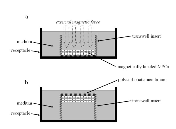
Magnetically labeled MSCs were plated at the bottom of an insert under the influence of an external magnetic force for 1 hour. Then the inserts were turned upside down for between 1 and 24 hours, and the number of MSCs which had fallen from the membrane was counted. The gene expression of MSCs affected magnetic force was analyzed with microarray. In the control group, the same procedure was done without the external magnetic force.
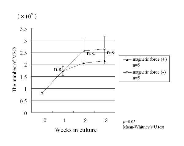
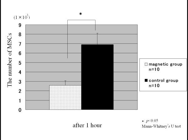
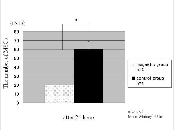
At 1 hour after the inserts were turned upside down, the average number of fallen MSCs in the magnetic group was significantly smaller than that in the control group, indicating enhanced cell adhesion. At 24 hours, the average number of fallen MSCs in the magnetic group was also significantly smaller than that in control group. In the magnetic group, integrin alpha2, alpha6, beta3 BP, intercellular adhesion molecule-2 (ICAM-2), platelet/endothelial cell adhesion molecule-1 (PECAM-1) were upregulated. At 1, 2 and 3 weeks after incubation, there was no statistical significant difference in the numbers of MSCs in the magnetic group and control group.
The results indicate that an external magnetic force for 1 hour enhances cell adhesion of MSCs. Moreover, there is no difference in cell proliferation after using an external magnetic force on magnetically labeled MSCs.
作为使用间充质干细胞(MSCs)进行移植的组织再生策略,有必要将MSCs聚集并保留在目标区域。为了有效聚集MSCs,我们开发了一种新型技术,即利用磁性标记的MSCs和外部磁力构建磁性靶向系统。在本研究中,我们从细胞黏附和增殖方面研究了外部磁力对磁性标记的MSCs的影响。
在外部磁力作用下,将磁性标记的MSCs接种于插入物底部1小时。然后将插入物倒置1至24小时,计数从膜上掉落的MSCs数量。用微阵列分析受磁力影响的MSCs的基因表达。对照组在无外部磁力的情况下进行相同操作。
插入物倒置1小时后,磁性组中掉落的MSCs平均数量显著少于对照组,表明细胞黏附增强。24小时时,磁性组中掉落的MSCs平均数量也显著少于对照组。在磁性组中,整合素α2、α6、β3 BP、细胞间黏附分子-2(ICAM-2)、血小板/内皮细胞黏附分子-1(PECAM-1)上调。培养1、2和3周后,磁性组和对照组的MSCs数量无统计学显著差异。
结果表明,1小时的外部磁力可增强MSCs的细胞黏附。此外,对磁性标记的MSCs施加外部磁力后,细胞增殖无差异。