• 文献检索
  • 文档翻译
  • 深度研究
  • 学术资讯
  • Suppr Zotero 插件Zotero 插件
  • 邀请有礼
  • 套餐&价格
  • 历史记录
应用&插件
Suppr Zotero 插件Zotero 插件浏览器插件Mac 客户端Windows 客户端微信小程序
定价
高级版会员购买积分包购买API积分包
服务
文献检索文档翻译深度研究API 文档MCP 服务
关于我们
关于 Suppr公司介绍联系我们用户协议隐私条款
关注我们

Suppr 超能文献

核心技术专利:CN118964589B侵权必究
粤ICP备2023148730 号-1Suppr @ 2026

文献检索

告别复杂PubMed语法,用中文像聊天一样搜索,搜遍4000万医学文献。AI智能推荐,让科研检索更轻松。

立即免费搜索

文件翻译

保留排版,准确专业,支持PDF/Word/PPT等文件格式,支持 12+语言互译。

免费翻译文档

深度研究

AI帮你快速写综述,25分钟生成高质量综述,智能提取关键信息,辅助科研写作。

立即免费体验

相似文献

1
Tumor targeting and imaging in live animals with functionalized semiconductor quantum rods.在活体动物中用功能化半导体量子点进行肿瘤靶向和成像。
ACS Appl Mater Interfaces. 2009 Mar;1(3):710-9. doi: 10.1021/am8002318.
2
Synthesis of cRGD-peptide conjugated near-infrared CdTe/ZnSe core-shell quantum dots for in vivo cancer targeting and imaging.cRGD 肽偶联近红外 CdTe/ZnSe 核壳量子点的合成及其用于体内癌症靶向和成像。
Chem Commun (Camb). 2010 Oct 14;46(38):7136-8. doi: 10.1039/c0cc00667j. Epub 2010 Aug 27.
3
Calixarene-coated water-soluble CdSe-ZnS semiconductor quantum dots that are highly fluorescent and stable in aqueous solution.杯芳烃包覆的水溶性CdSe-ZnS半导体量子点,在水溶液中具有高荧光性且稳定。
Chem Commun (Camb). 2005 Jun 14(22):2829-31. doi: 10.1039/b503178h. Epub 2005 Apr 21.
4
Biodistribution and toxicity assessment of intratumorally injected arginine-glycine-aspartic acid peptide conjugated to CdSe/ZnS quantum dots in mice bearing pancreatic neoplasm.Arg-Gly-Asp 肽偶联的 CdSe/ZnS 量子点瘤内注射在荷胰腺癌小鼠中的生物分布和毒性评估。
Chem Biol Interact. 2018 Aug 1;291:103-110. doi: 10.1016/j.cbi.2018.06.014. Epub 2018 Jun 14.
5
Investigation of biocompatible and protein sensitive highly luminescent quantum dots/nanocrystals of CdSe, CdSe/ZnS and CdSe/CdS.研究具有生物相容性和蛋白质敏感性的高亮度量子点/纳米晶体 CdSe、CdSe/ZnS 和 CdSe/CdS。
Spectrochim Acta A Mol Biomol Spectrosc. 2017 May 15;179:201-210. doi: 10.1016/j.saa.2017.02.028. Epub 2017 Feb 16.
6
CdSe/CdS/ZnS double shell nanorods with high photoluminescence efficiency and their exploitation as biolabeling probes.具有高光致发光效率的 CdSe/CdS/ZnS 双壳纳米棒及其作为生物标记探针的应用。
J Am Chem Soc. 2009 Mar 4;131(8):2948-58. doi: 10.1021/ja808369e.
7
Large enhancement of fluorescence efficiency from CdSe/ZnS quantum dots induced by resonant coupling to spatially controlled surface plasmons.通过与空间可控表面等离子体激元的共振耦合实现CdSe/ZnS量子点荧光效率的大幅增强。
Nano Lett. 2005 Aug;5(8):1557-61. doi: 10.1021/nl050813r.
8
Clustering of CdSe/CdS Quantum Dot/Quantum Rods into Micelles Can Form Bright, Non-blinking, Stable, and Biocompatible Probes.将CdSe/CdS量子点/量子棒聚集成胶束可形成明亮、不闪烁、稳定且具有生物相容性的探针。
Langmuir. 2015 Sep 1;31(34):9441-7. doi: 10.1021/acs.langmuir.5b01570. Epub 2015 Aug 18.
9
Toxicity assessment of repeated intravenous injections of arginine-glycine-aspartic acid peptide conjugated CdSeTe/ZnS quantum dots in mice.小鼠重复静脉注射精氨酸-甘氨酸-天冬氨酸肽偶联的CdSeTe/ZnS量子点的毒性评估
Int J Nanomedicine. 2014 Oct 17;9:4809-17. doi: 10.2147/IJN.S70092. eCollection 2014.
10
A robust and fast bacteria counting method using CdSe/ZnS core/shell quantum dots as labels.一种使用 CdSe/ZnS 核/壳量子点作为标记的稳健、快速的细菌计数方法。
J Microbiol Methods. 2009 Dec;79(3):367-70. doi: 10.1016/j.mimet.2009.09.019. Epub 2009 Sep 30.

引用本文的文献

1
Anisotropic nanomaterials for shape-dependent physicochemical and biomedical applications.各向异性纳米材料用于依赖形状的物理化学和生物医学应用。
Chem Soc Rev. 2019 Oct 7;48(19):5140-5176. doi: 10.1039/c9cs00011a. Epub 2019 Aug 29.
2
National Cancer Institute Alliance for nanotechnology in cancer-Catalyzing research and translation toward novel cancer diagnostics and therapeutics.美国国家癌症研究所癌症纳米技术联盟——推动癌症新型诊断和治疗的研究与转化。
Wiley Interdiscip Rev Nanomed Nanobiotechnol. 2019 Nov;11(6):e1570. doi: 10.1002/wnan.1570. Epub 2019 Jul 1.
3
Biocompatible near-infrared fluorescent nanoparticles for macro and microscopic in vivo functional bioimaging.用于体内宏观和微观功能生物成像的生物相容性近红外荧光纳米颗粒。
Biomed Opt Express. 2014 Oct 28;5(11):4076-88. doi: 10.1364/BOE.5.004076. eCollection 2014 Nov 1.
4
Targeted highly sensitive detection/eradication of multi-drug resistant Salmonella DT104 through gold nanoparticle-SWCNT bioconjugated nanohybrids.通过金纳米颗粒-单壁碳纳米管生物共轭纳米杂化物对多重耐药性肠炎沙门氏菌DT104进行靶向高灵敏度检测/根除。
Analyst. 2014 Aug 7;139(15):3702-5. doi: 10.1039/c4an00744a.
5
Photosensitive fluorescent dye contributes to phototoxicity and inflammatory responses of dye-doped silica NPs in cells and mice.光敏荧光染料会导致掺杂染料的二氧化硅纳米颗粒在细胞和小鼠中产生光毒性和炎症反应。
Theranostics. 2014 Feb 12;4(4):445-59. doi: 10.7150/thno.7653. eCollection 2014.
6
Quantum dot-based nanoprobes for in vivo targeted imaging.基于量子点的纳米探针用于体内靶向成像。
Curr Mol Med. 2013 Dec;13(10):1549-67. doi: 10.2174/1566524013666131111121733.
7
Quantum dots for cancer research: current status, remaining issues, and future perspectives.量子点在癌症研究中的应用:现状、遗留问题和未来展望。
Cancer Biol Med. 2012 Sep;9(3):151-63. doi: 10.7497/j.issn.2095-3941.2012.03.001.
8
Synthesis of luminescent near-infrared AgInS2 nanocrystals as optical probes for in vivo applications.合成具有近红外发光性能的 AgInS2 纳米晶体作为用于活体应用的光学探针。
Theranostics. 2013;3(2):109-15. doi: 10.7150/thno.5133. Epub 2013 Feb 1.
9
Image guided biodistribution and pharmacokinetic studies of theranostics.影像引导的治疗药物监测学的生物分布和药代动力学研究。
Theranostics. 2012;2(11):1040-53. doi: 10.7150/thno.4652. Epub 2012 Nov 5.
10
Bioconjugated pluronic triblock-copolymer micelle-encapsulated quantum dots for targeted imaging of cancer: in vitro and in vivo studies.用于癌症靶向成像的生物共轭普朗尼克三嵌段共聚物胶束包裹量子点:体外和体内研究
Theranostics. 2012;2(7):705-13. doi: 10.7150/thno.3456. Epub 2012 Jul 28.

本文引用的文献

1
Bio-conjugated luminescent quantum dots of doped ZnS: a cyto-friendly system for targeted cancer imaging.生物共轭的掺杂硫化锌发光量子点:一种用于靶向癌症成像的细胞友好型系统。
Nanotechnology. 2009 Feb 11;20(6):065102. doi: 10.1088/0957-4484/20/6/065102. Epub 2009 Jan 14.
2
Mn-doped near-infrared quantum dots as multimodal targeted probes for pancreatic cancer imaging.锰掺杂近红外量子点作为胰腺癌成像的多模态靶向探针
Nanotechnology. 2009 Jan 7;20(1):015102. doi: 10.1088/0957-4484/20/1/015102. Epub 2008 Dec 5.
3
Composite nanoparticles take aim at cancer.复合纳米颗粒瞄准癌症。
ACS Nano. 2008 Nov 25;2(11):2200-5. doi: 10.1021/nn800716j.
4
An orally bioavailable small-molecule inhibitor of Hedgehog signaling inhibits tumor initiation and metastasis in pancreatic cancer.一种口服生物可利用的刺猬信号通路小分子抑制剂可抑制胰腺癌的肿瘤起始和转移。
Mol Cancer Ther. 2008 Sep;7(9):2725-35. doi: 10.1158/1535-7163.MCT-08-0573.
5
In vivo real-time bioimaging of hyaluronic acid derivatives using quantum dots.使用量子点对透明质酸衍生物进行体内实时生物成像。
Biopolymers. 2008 Dec;89(12):1144-53. doi: 10.1002/bip.21066.
6
Bioconjugated quantum rods as targeted probes for efficient transmigration across an in vitro blood-brain barrier.生物共轭量子棒作为高效穿越体外血脑屏障的靶向探针。
Bioconjug Chem. 2008 Jun;19(6):1179-85. doi: 10.1021/bc700477u. Epub 2008 May 13.
7
Non-invasive near infrared fluorescence imaging of CdHgTe quantum dots in mouse model.小鼠模型中碲镉汞量子点的无创近红外荧光成像
J Fluoresc. 2008 Sep;18(5):801-11. doi: 10.1007/s10895-007-0307-9. Epub 2008 Jan 5.
8
In vivo tumor targeting and spectroscopic detection with surface-enhanced Raman nanoparticle tags.利用表面增强拉曼纳米颗粒标签进行体内肿瘤靶向和光谱检测。
Nat Biotechnol. 2008 Jan;26(1):83-90. doi: 10.1038/nbt1377. Epub 2007 Dec 23.
9
Compact cysteine-coated CdSe(ZnCdS) quantum dots for in vivo applications.用于体内应用的紧凑型半胱氨酸包覆的CdSe(ZnCdS)量子点。
J Am Chem Soc. 2007 Nov 28;129(47):14530-1. doi: 10.1021/ja073790m. Epub 2007 Nov 6.
10
Renal clearance of quantum dots.量子点的肾脏清除率。
Nat Biotechnol. 2007 Oct;25(10):1165-70. doi: 10.1038/nbt1340. Epub 2007 Sep 23.

在活体动物中用功能化半导体量子点进行肿瘤靶向和成像。

Tumor targeting and imaging in live animals with functionalized semiconductor quantum rods.

机构信息

Institute for Lasers, Photonics and Biophotonics, The State University of New York at Buffalo, Buffalo, NY 14260-4200, USA.

出版信息

ACS Appl Mater Interfaces. 2009 Mar;1(3):710-9. doi: 10.1021/am8002318.

DOI:10.1021/am8002318
PMID:20160901
原文链接:https://pmc.ncbi.nlm.nih.gov/articles/PMC2768400/
Abstract

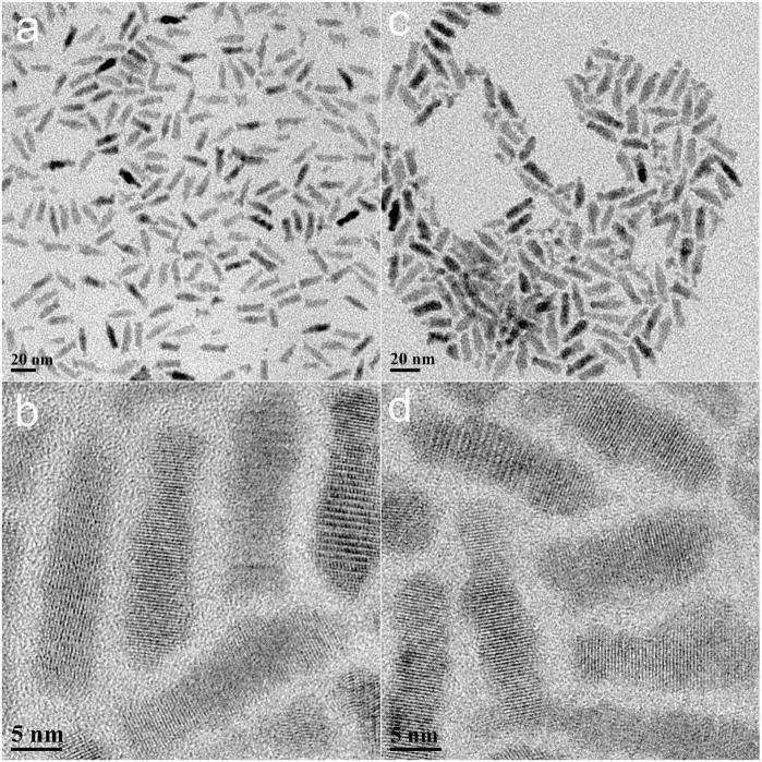
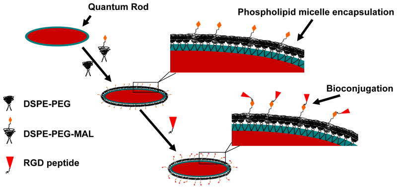
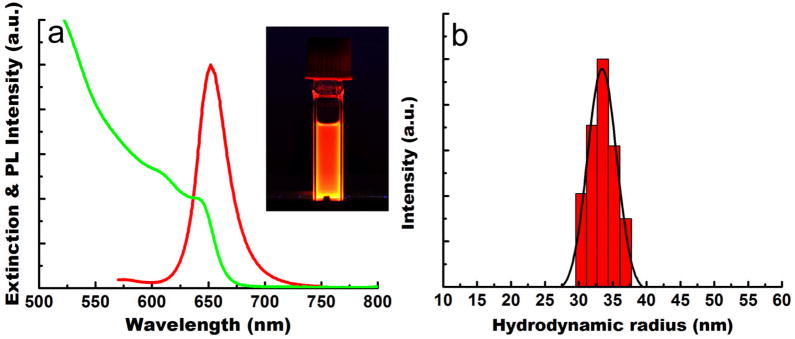
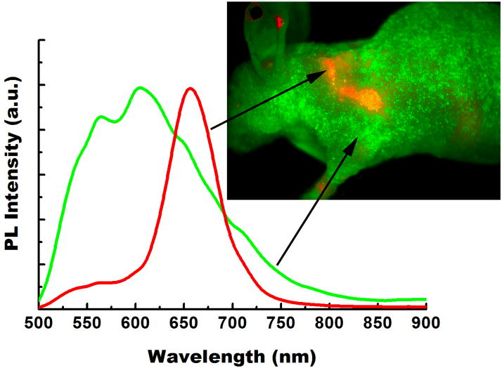
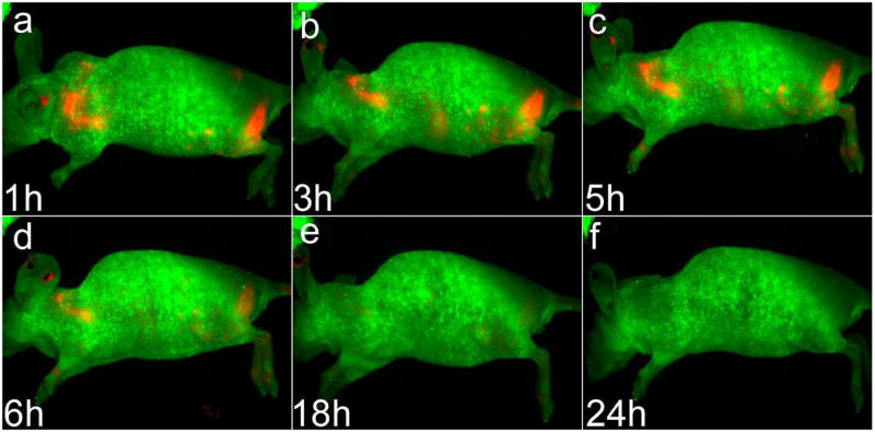
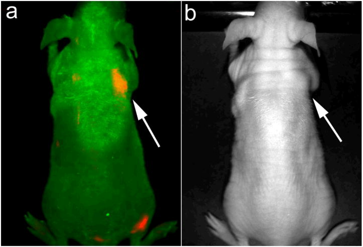
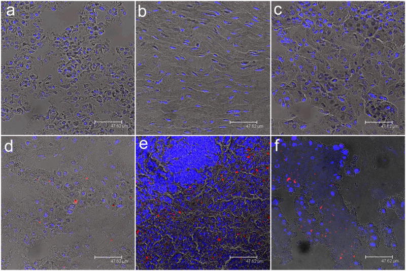
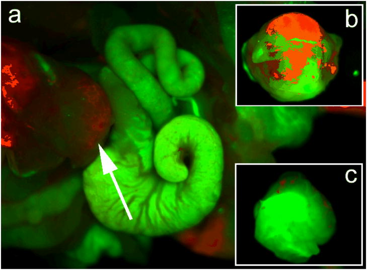
In this contribution, we demonstrate that highly luminescent CdSe/CdS/ZnS quantum rods (QRs) coated with PEGylated phospholipids and conjugated with cyclic RGD peptide can be successfully used for tumor targeting and imaging in live animals. The design of these targeted luminescent probes involves encapsulation of hydrophobic CdSe/CdS/ZnS QRs with PEGylated phospholipids, followed by conjugation of these PEGylated phospholipids to ligands that specifically target the tumor vasculature. In vivo optical imaging studies in nude mice bearing pancreatic cancer xenografts, both subcutaneous and orthotopic, indicate that the QR probes accumulate at tumor sites via the cyclic RGD peptides on the QR surface binding to the alpha(V)beta(3) integrins overexpressed in the tumor vasculature, following systemic injection. In vivo tumor detection studies showed no adverse effects even at a dose roughly 6.5 times higher than has been reported for in vivo imaging studies using quantum dots. Cytotoxicity studies indicated the absence of any toxic effect in the cellular and tissue levels arising from functionalized QRs. These results demonstrate the vast potential of QRs as bright, photostable, and biocompatible luminescent probes for the early diagnosis of cancer.

摘要

在本研究中,我们证明了经聚乙二醇化磷脂和环状 RGD 肽修饰的具有高光致发光性能的 CdSe/CdS/ZnS 量子点(QRs)可成功用于活体动物中的肿瘤靶向和成像。这些靶向发光探针的设计涉及将疏水性 CdSe/CdS/ZnS QRs 用聚乙二醇化磷脂包封,然后将这些聚乙二醇化磷脂与专门针对肿瘤血管系统的配体偶联。在携带胰腺癌细胞异种移植物的裸鼠(皮下和原位)的体内光学成像研究中,表明 QR 探针通过 QR 表面上的环状 RGD 肽与肿瘤血管系统中过度表达的 α(V)β(3)整合素结合,在系统注射后在肿瘤部位积累。体内肿瘤检测研究表明,即使在大约比使用量子点进行体内成像研究报道的剂量高 6.5 倍的剂量下,也没有任何不良反应。细胞毒性研究表明,功能性化 QR 不会在细胞和组织水平上产生任何毒性作用。这些结果表明 QR 作为明亮、光稳定且生物相容的发光探针在癌症的早期诊断中具有巨大的潜力。

https://cdn.ncbi.nlm.nih.gov/pmc/blobs/3091/2768400/02ebccfb2a1f/nihms110432f9.jpg
https://cdn.ncbi.nlm.nih.gov/pmc/blobs/3091/2768400/645442af3bb2/nihms110432f1.jpg
https://cdn.ncbi.nlm.nih.gov/pmc/blobs/3091/2768400/b254df76caeb/nihms110432f2.jpg
https://cdn.ncbi.nlm.nih.gov/pmc/blobs/3091/2768400/f5fb4b32f448/nihms110432f3.jpg
https://cdn.ncbi.nlm.nih.gov/pmc/blobs/3091/2768400/ff7b0cbb5513/nihms110432f4.jpg
https://cdn.ncbi.nlm.nih.gov/pmc/blobs/3091/2768400/0d49a838469b/nihms110432f5.jpg
https://cdn.ncbi.nlm.nih.gov/pmc/blobs/3091/2768400/0ae794e315c7/nihms110432f6.jpg
https://cdn.ncbi.nlm.nih.gov/pmc/blobs/3091/2768400/adab0960c30e/nihms110432f7.jpg
https://cdn.ncbi.nlm.nih.gov/pmc/blobs/3091/2768400/409a19f901fe/nihms110432f8.jpg
https://cdn.ncbi.nlm.nih.gov/pmc/blobs/3091/2768400/02ebccfb2a1f/nihms110432f9.jpg