Advanced Light Microscopy Core Facility, European Molecular Biology Laboratory Heidelberg, D-69117 Heidelberg, Germany.
J Cell Biol. 2010 Feb 22;188(4):453-61. doi: 10.1083/jcb.200910105.
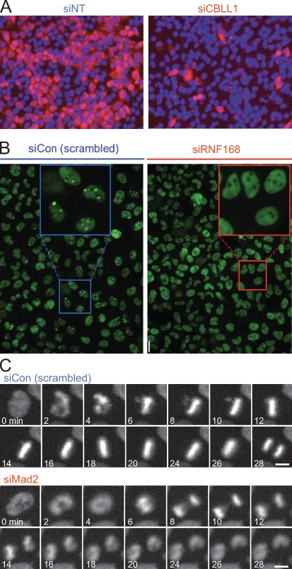
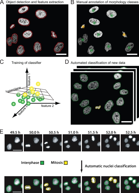
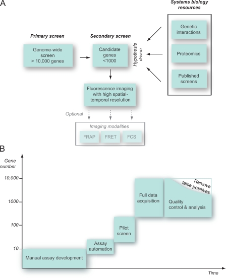
Fluorescence microscopy is one of the most powerful tools to investigate complex cellular processes such as cell division, cell motility, or intracellular trafficking. The availability of RNA interference (RNAi) technology and automated microscopy has opened the possibility to perform cellular imaging in functional genomics and other large-scale applications. Although imaging often dramatically increases the content of a screening assay, it poses new challenges to achieve accurate quantitative annotation and therefore needs to be carefully adjusted to the specific needs of individual screening applications. In this review, we discuss principles of assay design, large-scale RNAi, microscope automation, and computational data analysis. We highlight strategies for imaging-based RNAi screening adapted to different library and assay designs.
荧光显微镜是研究细胞分裂、细胞运动或细胞内运输等复杂细胞过程的最有力工具之一。RNA 干扰 (RNAi) 技术和自动化显微镜的可用性为功能基因组学和其他大规模应用中的细胞成像开辟了可能性。虽然成像通常会极大地增加筛选测定的信息量,但它给实现准确的定量注释带来了新的挑战,因此需要根据个别筛选应用的具体需求进行仔细调整。在这篇综述中,我们讨论了测定设计、大规模 RNAi、显微镜自动化和计算数据分析的原理。我们强调了适用于不同文库和测定设计的基于成像的 RNAi 筛选的策略。