Laboratory for Spectral Diagnosis, Department of Chemistry and Chemical Biology, Northeastern University, Boston, MA 02115, USA.
Lab Invest. 2010 Jul;90(7):1068-77. doi: 10.1038/labinvest.2010.72. Epub 2010 Apr 5.
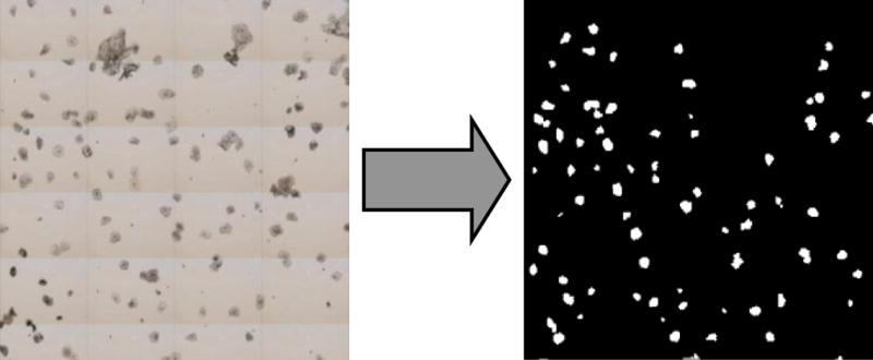
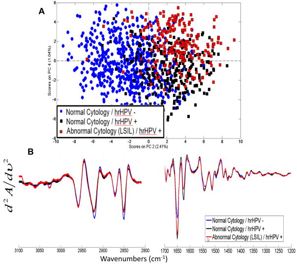
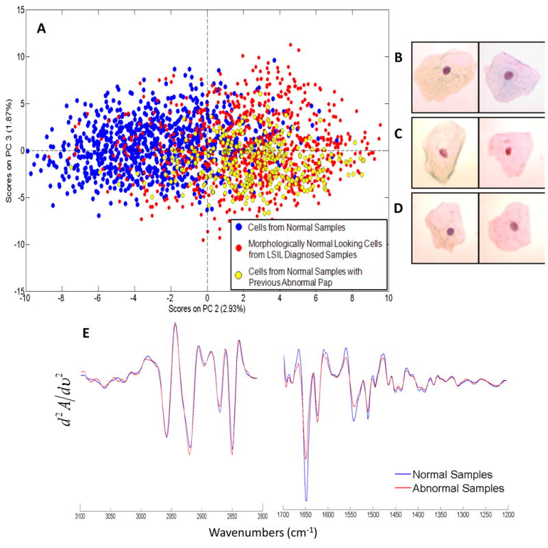
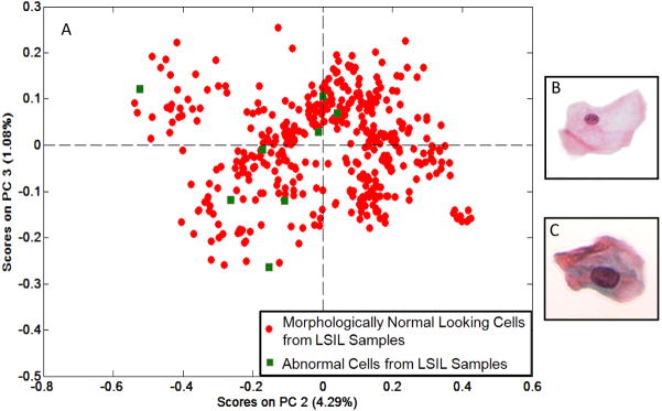
Spectral cytopathology (SCP) is a novel spectroscopic method for objective and unsupervised classification of individual exfoliated cells. The limitations of conventional cytopathology are well recognized within the pathology community. In SCP, cellular differentiation is made by observing molecular changes in the nucleus and the cytoplasm, which may or may not produce morphological changes detectable by conventional cytopathology. This proof of concept study shows SCP's potential as an enhancing tool for cytopathologists by aiding in the accurate and reproducible diagnosis of cells in all states of disease. Infrared spectra are collected from cervical cells deposited onto reflectively coated glass slides. Each cell has a corresponding infrared spectrum that describes its unique biochemical composition. Spectral data are processed and analyzed by an unsupervised chemometric algorithm, principal component analysis. In this blind study, cervical samples are classified by analyzing the spectra of morphologically normal looking squamous cells from normal samples and samples diagnosed by conventional cytopathology with low-grade squamous intraepithelial lesions. SCP discriminated cytopathological diagnoses amongst 12 different cervical samples with a high degree of specificity and sensitivity. SCP also correlated two samples with abnormal spectral changes: these samples had a normal cytopathological diagnosis but had a history of abnormal cervical cytology. The spectral changes observed in the morphologically normal looking cells are most likely because of an infection with human papillomavirus (HPV). HPV DNA testing was conducted on five additional samples, and SCP accurately differentiated these samples by their HPV status. SCP tracks biochemical variations in cells that are consistent with the onset of disease. HPV has been implicated as the cause of these changes detected spectroscopically. SCP does not depend on identifying the sparse number of morphologically abnormal cells within a large sample to make an accurate classification, as does conventional cytopathology. These findings suggest that the detection of cellular biochemical variations by SCP can serve as a new enhancing screening method that can identify earlier stages of disease.
光谱细胞病理学(SCP)是一种新颖的光谱方法,可用于对单个脱落细胞进行客观的、无监督的分类。传统细胞病理学的局限性在病理学领域是众所周知的。在 SCP 中,通过观察细胞核和细胞质中的分子变化来区分细胞的分化,这些变化可能会产生也可能不会产生传统细胞病理学可检测到的形态变化。这项概念验证研究表明,SCP 通过帮助准确和可重复地诊断处于疾病各个阶段的细胞,具有作为细胞病理学家增强工具的潜力。从沉积在反射性涂层玻片上的宫颈细胞中收集红外光谱。每个细胞都有一个对应的红外光谱,描述了其独特的生化组成。通过无监督化学计量算法(主成分分析)处理和分析光谱数据。在这项盲法研究中,通过分析来自正常样本和经传统细胞病理学诊断为低级别鳞状上皮内病变的样本的形态正常的鳞状细胞的光谱,对宫颈样本进行分类。SCP 以高度的特异性和敏感性区分了 12 个不同的宫颈样本的细胞病理诊断。SCP 还将两个具有异常光谱变化的样本进行了分类:这些样本的细胞病理诊断正常,但宫颈细胞学异常史。在形态正常的细胞中观察到的光谱变化很可能是由于感染了人乳头瘤病毒(HPV)。对另外五个样本进行了 HPV DNA 检测,SCP 准确地根据 HPV 状态对这些样本进行了区分。SCP 跟踪了与疾病发生一致的细胞生化变化。HPV 被认为是光谱检测到的这些变化的原因。SCP 不依赖于在大量样本中识别少数形态异常的细胞来进行准确的分类,这与传统细胞病理学不同。这些发现表明,SCP 可以检测细胞生化变化,作为一种新的增强筛选方法,可以识别疾病的早期阶段。