Department of Pediatrics, Yale University School of Medicine, New Haven, CT, USA.
Nutr Metab (Lond). 2010 May 11;7:41. doi: 10.1186/1743-7075-7-41.
To compare the relationship of skeletal muscle mass with bone mineral content in an ethnically diverse group of 6 to 18 year old boys and girls.
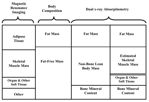
175 healthy children (103 boys; 72 girls) had assessments of body mass, height, and Tanner stage. Whole body bone mineral content, non-bone lean body mass (nbLBM), skeletal muscle mass, and fat mass were assessed using dual-energy X-ray absorptiometry (DXA). Muscle mass was estimated from an equation using appendicular lean soft tissue measured by DXA, weight and height. Estimates of skeletal muscle mass and adipose tissue were also assessed by whole body multi-slice magnetic resonance imaging (MRI). Linear regression was used to determine whether skeletal muscle mass assessed by DXA or by MRI were better predictors of bone mineral content compared with nbLBM after adjusting for sex, age, race or ethnicity, and Tanner stage.
Greater skeletal muscle mass was associated with greater bone mineral content (p < 0.001). The skeletal muscle mass assessed by MRI provided a better fitting regression model (determined by R2 statistic) compared with assessment by DXA for predicting bone mineral content. The proportion of skeletal muscle mass in nbLBM was significantly associated with greater bone mineral content adjusted for total nbLBM.
This study is among the first to describe and compare the relationship of skeletal muscle to bone using both MRI and DXA estimates. The results demonstrate that the use of MRI provides a modestly better fitting model for the relationship of skeletal muscle to bone compared with DXA. Skeletal muscle had an impact on bone mineral content independent of total non-bone lean body mass. In addition, Hispanics had greater bone mineral content compared to other race and ethnic groups after adjusting for sex, age, adipose tissue, skeletal muscle mass, and height.
为了比较种族多样化的 6 至 18 岁男孩和女孩群体中骨骼肌量与骨矿物质含量的关系。
175 名健康儿童(103 名男孩;72 名女孩)接受了体重、身高和 Tanner 分期的评估。使用双能 X 射线吸收法(DXA)评估全身骨矿物质含量、非骨瘦体重(nbLBM)、骨骼肌量和脂肪量。肌肉量通过 DXA 测量的四肢瘦软组织、体重和身高的方程进行估算。还通过全身多切片磁共振成像(MRI)评估骨骼肌量和脂肪组织的估计值。线性回归用于确定在调整性别、年龄、种族或民族以及 Tanner 分期后,通过 DXA 或 MRI 评估的骨骼肌量与 nbLBM 相比,哪个更能预测骨矿物质含量。
骨骼肌量越大,骨矿物质含量越高(p<0.001)。与 DXA 评估相比,MRI 评估的骨骼肌量提供了更好的拟合回归模型(由 R2 统计量确定),用于预测骨矿物质含量。在调整总 nbLBM 后,骨骼肌量在 nbLBM 中的比例与更大的骨矿物质含量显著相关。
本研究首次使用 MRI 和 DXA 评估来描述和比较骨骼肌与骨骼的关系。结果表明,与 DXA 相比,MRI 的使用为骨骼肌与骨骼的关系提供了一个稍好的拟合模型。骨骼肌对骨矿物质含量的影响独立于总非骨瘦体重。此外,在调整性别、年龄、脂肪组织、骨骼肌量和身高后,西班牙裔的骨矿物质含量高于其他种族和民族群体。