• 文献检索
  • 文档翻译
  • 深度研究
  • 学术资讯
  • Suppr Zotero 插件Zotero 插件
  • 邀请有礼
  • 套餐&价格
  • 历史记录
应用&插件
Suppr Zotero 插件Zotero 插件浏览器插件Mac 客户端Windows 客户端微信小程序
定价
高级版会员购买积分包购买API积分包
服务
文献检索文档翻译深度研究API 文档MCP 服务
关于我们
关于 Suppr公司介绍联系我们用户协议隐私条款
关注我们

Suppr 超能文献

核心技术专利:CN118964589B侵权必究
粤ICP备2023148730 号-1Suppr @ 2026

文献检索

告别复杂PubMed语法,用中文像聊天一样搜索,搜遍4000万医学文献。AI智能推荐,让科研检索更轻松。

立即免费搜索

文件翻译

保留排版,准确专业,支持PDF/Word/PPT等文件格式,支持 12+语言互译。

免费翻译文档

深度研究

AI帮你快速写综述,25分钟生成高质量综述,智能提取关键信息,辅助科研写作。

立即免费体验

前列腺 MRI 与 3D MR 波谱:我们的做法。

Prostate MRI and 3D MR spectroscopy: how we do it.

机构信息

Department of Radiology, University of Cincinnati Medical Center, Cincinnati, OH 45267-0761, USA.

出版信息

AJR Am J Roentgenol. 2010 Jun;194(6):1414-26. doi: 10.2214/AJR.10.4312.

DOI:10.2214/AJR.10.4312
PMID:20489079
原文链接:https://pmc.ncbi.nlm.nih.gov/articles/PMC2895419/
Abstract

OBJECTIVE

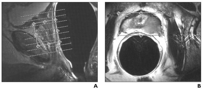
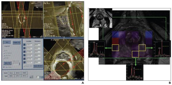
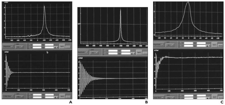
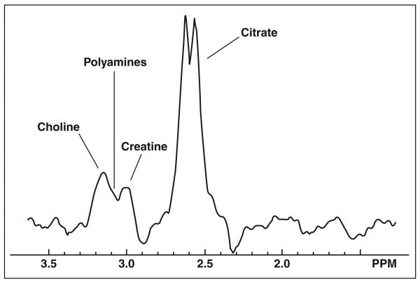
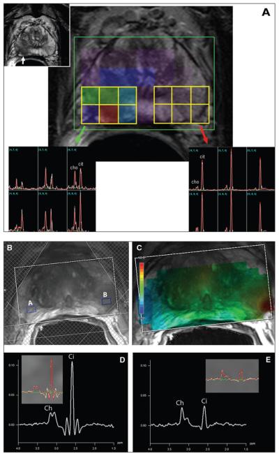
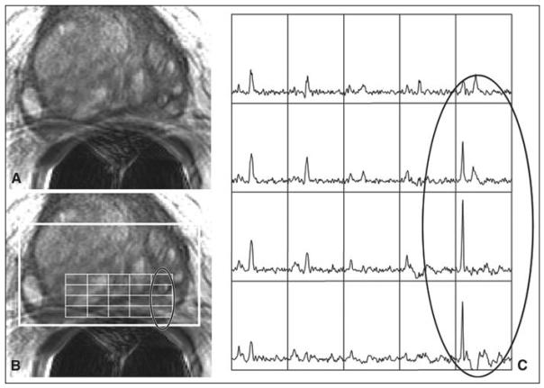
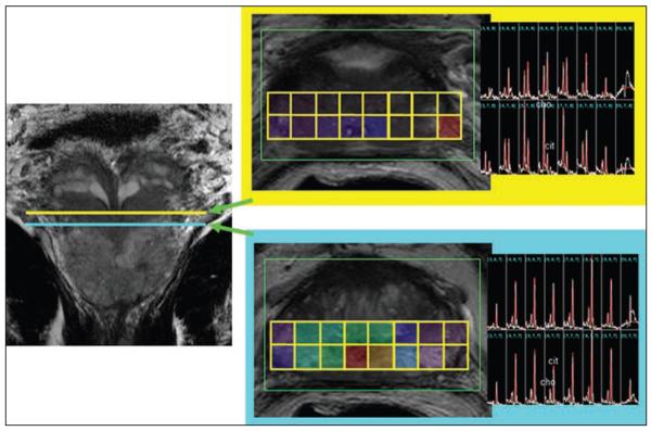
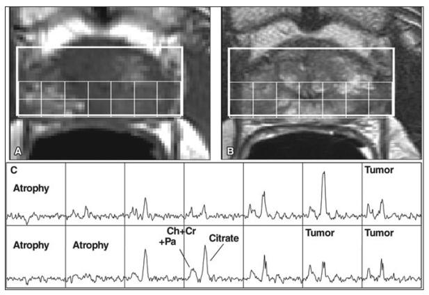
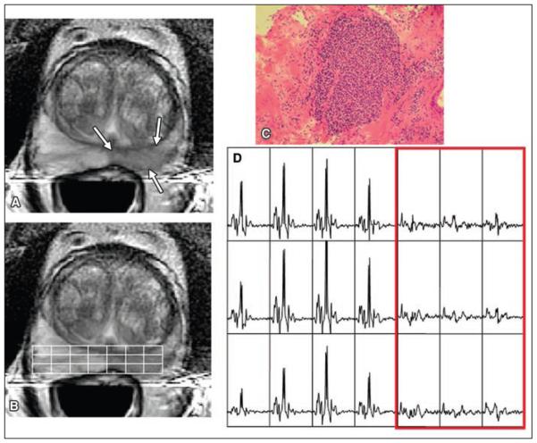
This review is a primer on the technical aspects of performing a high-quality MRI and MR spectroscopic imaging examination of the prostate.

CONCLUSION

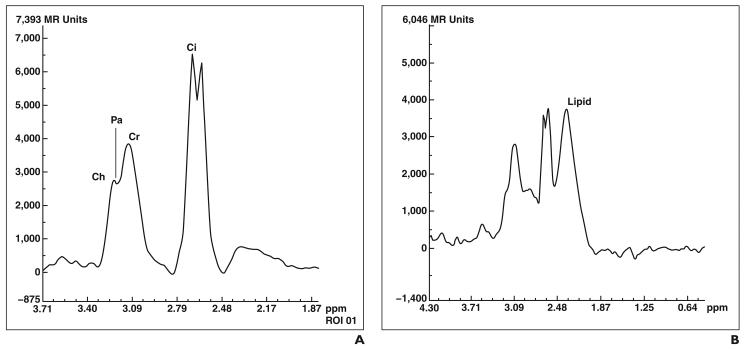
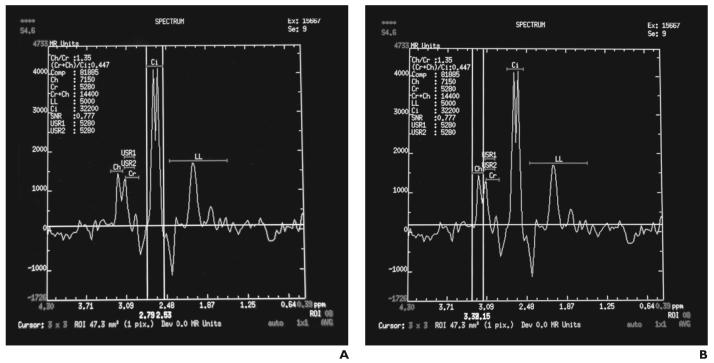
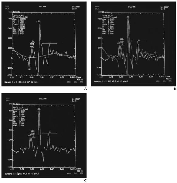
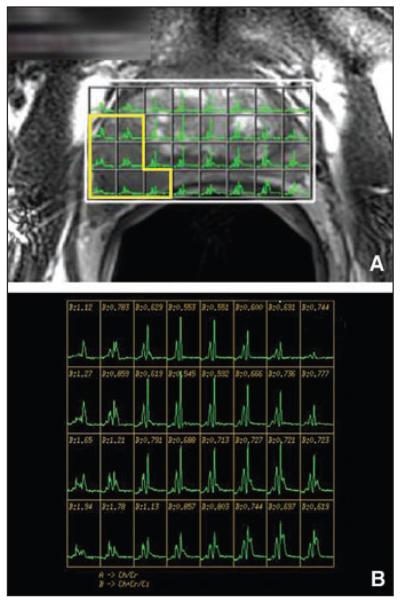
MRI and MR spectroscopic imaging are useful tools in the localization, staging, and functional assessment of prostate cancer. Performing a high-quality MR spectroscopic examination requires understanding of the technical aspects and limitations of spectral acquisition, postprocessing techniques, and spectral evaluation.

摘要

目的

本综述是关于前列腺进行高质量 MRI 和 MR 波谱成像检查的技术方面的入门介绍。

结论

MRI 和 MR 波谱成像是前列腺癌定位、分期和功能评估的有用工具。进行高质量的 MR 波谱检查需要了解光谱采集、后处理技术和光谱评估的技术方面和局限性。

https://cdn.ncbi.nlm.nih.gov/pmc/blobs/bbba/2895419/7b8f78d35d5b/nihms-209138-f0018.jpg
https://cdn.ncbi.nlm.nih.gov/pmc/blobs/bbba/2895419/45a0bb8d9d6e/nihms-209138-f0001.jpg
https://cdn.ncbi.nlm.nih.gov/pmc/blobs/bbba/2895419/fa037e6a002f/nihms-209138-f0002.jpg
https://cdn.ncbi.nlm.nih.gov/pmc/blobs/bbba/2895419/d9d73477baf9/nihms-209138-f0003.jpg
https://cdn.ncbi.nlm.nih.gov/pmc/blobs/bbba/2895419/0b6ed6a0b245/nihms-209138-f0004.jpg
https://cdn.ncbi.nlm.nih.gov/pmc/blobs/bbba/2895419/860982208790/nihms-209138-f0005.jpg
https://cdn.ncbi.nlm.nih.gov/pmc/blobs/bbba/2895419/ccf3954be103/nihms-209138-f0006.jpg
https://cdn.ncbi.nlm.nih.gov/pmc/blobs/bbba/2895419/aea3a733df4b/nihms-209138-f0007.jpg
https://cdn.ncbi.nlm.nih.gov/pmc/blobs/bbba/2895419/2084cf955aab/nihms-209138-f0008.jpg
https://cdn.ncbi.nlm.nih.gov/pmc/blobs/bbba/2895419/8bb699e78796/nihms-209138-f0009.jpg
https://cdn.ncbi.nlm.nih.gov/pmc/blobs/bbba/2895419/1f0dbcb639b0/nihms-209138-f0010.jpg
https://cdn.ncbi.nlm.nih.gov/pmc/blobs/bbba/2895419/20b02099a572/nihms-209138-f0011.jpg
https://cdn.ncbi.nlm.nih.gov/pmc/blobs/bbba/2895419/9d5c7d28519a/nihms-209138-f0012.jpg
https://cdn.ncbi.nlm.nih.gov/pmc/blobs/bbba/2895419/4ffdfcf5820f/nihms-209138-f0013.jpg
https://cdn.ncbi.nlm.nih.gov/pmc/blobs/bbba/2895419/281723f49a41/nihms-209138-f0014.jpg
https://cdn.ncbi.nlm.nih.gov/pmc/blobs/bbba/2895419/e8296835ce1f/nihms-209138-f0015.jpg
https://cdn.ncbi.nlm.nih.gov/pmc/blobs/bbba/2895419/55d28781851a/nihms-209138-f0016.jpg
https://cdn.ncbi.nlm.nih.gov/pmc/blobs/bbba/2895419/07949a6c42b9/nihms-209138-f0017.jpg
https://cdn.ncbi.nlm.nih.gov/pmc/blobs/bbba/2895419/7b8f78d35d5b/nihms-209138-f0018.jpg

相似文献

1
Prostate MRI and 3D MR spectroscopy: how we do it.前列腺 MRI 与 3D MR 波谱:我们的做法。
AJR Am J Roentgenol. 2010 Jun;194(6):1414-26. doi: 10.2214/AJR.10.4312.
2
[Comparison of the accuracy of manual and automatic evaluation of MR spectra in prostate carcinoma].
Urologe A. 2007 Sep;46(9):1252. doi: 10.1007/s00120-007-1488-1.
3
Magnetic resonance spectroscopic imaging of benign prostatic tissue: findings at 3.0 T compared to 1.5 T-initial experience.良性前列腺组织的磁共振波谱成像:3.0T 与 1.5T 的比较——初步经验。
Clin Imaging. 2011 Jul-Aug;35(4):288-93. doi: 10.1016/j.clinimag.2010.07.005.
4
[MR spectroscopic imaging for evaluation of prostate cancer].[磁共振波谱成像在前列腺癌评估中的应用]
J Radiol. 2006 Feb;87(2 Pt 2):222-7. doi: 10.1016/s0221-0363(06)73996-8.
5
Prostate cancer: metabolic response to cryosurgery as detected with 3D H-1 MR spectroscopic imaging.前列腺癌:通过三维氢-1磁共振波谱成像检测到的对冷冻手术的代谢反应。
Radiology. 1996 Aug;200(2):489-96. doi: 10.1148/radiology.200.2.8685346.
6
[Multiparametric magnetic resonance imaging of the prostate - technique and clinical applications].[前列腺多参数磁共振成像——技术与临床应用]
Rofo. 2011 Jul;183(7):607-17. doi: 10.1055/s-0029-1246055. Epub 2011 Apr 12.
7
MR imaging and MR spectroscopy in prostate cancer management.磁共振成像和磁共振波谱在前列腺癌管理中的应用
Radiol Clin North Am. 2006 Sep;44(5):723-34, viii. doi: 10.1016/j.rcl.2006.07.008.
8
Proton MR spectroscopy of the prostate.前列腺的质子磁共振波谱分析。
Eur J Radiol. 2007 Sep;63(3):351-60. doi: 10.1016/j.ejrad.2007.06.024. Epub 2007 Aug 20.
9
Metabolite ratios in 1H MR spectroscopic imaging of the prostate.前列腺1H磁共振波谱成像中的代谢物比率
Magn Reson Med. 2015 Jan;73(1):1-12. doi: 10.1002/mrm.25122. Epub 2014 Jan 31.
10
[1H magnetic resonance spectroscopy of the prostate].[前列腺的1H磁共振波谱分析]
Radiologe. 2003 Jun;43(6):481-8. doi: 10.1007/s00117-003-0902-y.

引用本文的文献

1
Integration of magnetic resonance imaging and deep learning for prostate cancer detection: a systematic review.磁共振成像与深度学习整合用于前列腺癌检测的系统评价
Am J Clin Exp Urol. 2025 Apr 25;13(2):69-91. doi: 10.62347/CSIJ8326. eCollection 2025.
2
New imaging techniques and trends in radiology.放射学中的新成像技术与趋势
Diagn Interv Radiol. 2025 Jan 16. doi: 10.4274/dir.2024.242926.
3
Developments in proton MR spectroscopic imaging of prostate cancer.前列腺癌质子磁共振波谱成像的研究进展。
MAGMA. 2022 Aug;35(4):645-665. doi: 10.1007/s10334-022-01011-9. Epub 2022 Apr 20.
4
Role of magnetic resonance spectroscopy in differential diagnosis of solitary pulmonary lesions.磁共振波谱在肺部单发占位性病变鉴别诊断中的作用。
Diagn Interv Radiol. 2021 Nov;27(6):710-715. doi: 10.5152/dir.2021.20419.
5
Positron Emission Tomography Imaging of Prostate Cancer with Ga-68-Labeled Gastrin-Releasing Peptide Receptor Agonist BBN and Antagonist RM26.镓-68 标记胃泌素释放肽受体激动剂 BBN 和拮抗剂 RM26 用于前列腺癌的正电子发射断层成像
Bioconjug Chem. 2018 Feb 21;29(2):410-419. doi: 10.1021/acs.bioconjchem.7b00726. Epub 2018 Jan 9.
6
Metabolic Vulnerabilities of Prostate Cancer: Diagnostic and Therapeutic Opportunities.前列腺癌的代谢脆弱性:诊断和治疗机会。
Cold Spring Harb Perspect Med. 2018 Oct 1;8(10):a030569. doi: 10.1101/cshperspect.a030569.
7
Impact of the integration of proton magnetic resonance imaging spectroscopy to PI-RADS 2 for prediction of high grade and high stage prostate cancer.将质子磁共振成像光谱整合到PI-RADS 2中对预测高级别和高分期前列腺癌的影响。
Radiol Bras. 2017 Sep-Oct;50(5):299-307. doi: 10.1590/0100-3984.2016.0117.
8
Proton magnetic resonance spectroscopy in oncology: the fingerprints of cancer?肿瘤学中的质子磁共振波谱:癌症的指纹图谱?
Diagn Interv Radiol. 2016 Jan-Feb;22(1):75-89. doi: 10.5152/dir.2015.15009.
9
Integration of tissue metabolomics, transcriptomics and immunohistochemistry reveals ERG- and gleason score-specific metabolomic alterations in prostate cancer.组织代谢组学、转录组学和免疫组化的整合揭示了前列腺癌中ERG和Gleason评分特异性的代谢组学改变。
Oncotarget. 2016 Jan 12;7(2):1421-38. doi: 10.18632/oncotarget.6370.
10
The value of prostate MRI with endorectal coil in detecting seminal vesicle involvement in patients with prostate cancer.经直肠线圈前列腺MRI在检测前列腺癌患者精囊受累情况中的价值。
Iran J Radiol. 2015 Jan 17;12(1):e14556. doi: 10.5812/iranjradiol.14556. eCollection 2015 Jan.

本文引用的文献

1
Mucinous adenocarcinoma of the prostate: MRI and MR spectroscopy features.前列腺黏液腺癌:MRI及磁共振波谱特征
AJR Am J Roentgenol. 2009 Sep;193(3):W238-43. doi: 10.2214/AJR.08.1495.
2
Advances in MR spectroscopy of the prostate.前列腺磁共振波谱成像的进展
Magn Reson Imaging Clin N Am. 2008 Nov;16(4):697-710, ix-x. doi: 10.1016/j.mric.2008.07.005.
3
Three-dimensional proton MR spectroscopy of human prostate at 3 T without endorectal coil: feasibility.3T下无直肠内线圈的人体前列腺三维质子磁共振波谱分析:可行性
Radiology. 2007 Nov;245(2):507-16. doi: 10.1148/radiol.2451061444. Epub 2007 Sep 11.
4
High-speed 3T MR spectroscopic imaging of prostate with flyback echo-planar encoding.采用回波平面编码的前列腺高速3T磁共振波谱成像
J Magn Reson Imaging. 2007 Jun;25(6):1288-92. doi: 10.1002/jmri.20916.
5
Prostate cancer: body-array versus endorectal coil MR imaging at 3 T--comparison of image quality, localization, and staging performance.前列腺癌:3T 下体部阵列与直肠内线圈磁共振成像——图像质量、定位及分期性能的比较
Radiology. 2007 Jul;244(1):184-95. doi: 10.1148/radiol.2441060425. Epub 2007 May 10.
6
Standardized threshold approach using three-dimensional proton magnetic resonance spectroscopic imaging in prostate cancer localization of the entire prostate.在整个前列腺癌定位中使用三维质子磁共振波谱成像的标准化阈值方法。
Invest Radiol. 2007 Feb;42(2):116-22. doi: 10.1097/01.rli.0000251541.03822.bb.
7
MR imaging and MR spectroscopic imaging in the pre-treatment evaluation of prostate cancer.磁共振成像和磁共振波谱成像在前列腺癌治疗前评估中的应用
Br J Radiol. 2005;78 Spec No 2:S103-11. doi: 10.1259/bjr/11253478.
8
Optimal timing for in vivo 1H-MR spectroscopic imaging of the human prostate at 3T.3T下人体前列腺活体1H磁共振波谱成像的最佳时机
Magn Reson Med. 2005 Jun;53(6):1268-74. doi: 10.1002/mrm.20468.
9
Sequence design for magnetic resonance spectroscopic imaging of prostate cancer at 3 T.3T 下前列腺癌磁共振波谱成像的序列设计
Magn Reson Med. 2005 May;53(5):1033-9. doi: 10.1002/mrm.20478.
10
Prostate depiction at endorectal MR spectroscopic imaging: investigation of a standardized evaluation system.直肠内磁共振波谱成像对前列腺的描绘:一种标准化评估系统的研究
Radiology. 2004 Dec;233(3):701-8. doi: 10.1148/radiol.2333030672.