• 文献检索
  • 文档翻译
  • 深度研究
  • 学术资讯
  • Suppr Zotero 插件Zotero 插件
  • 邀请有礼
  • 套餐&价格
  • 历史记录
应用&插件
Suppr Zotero 插件Zotero 插件浏览器插件Mac 客户端Windows 客户端微信小程序
定价
高级版会员购买积分包购买API积分包
服务
文献检索文档翻译深度研究API 文档MCP 服务
关于我们
关于 Suppr公司介绍联系我们用户协议隐私条款
关注我们

Suppr 超能文献

核心技术专利:CN118964589B侵权必究
粤ICP备2023148730 号-1Suppr @ 2026

文献检索

告别复杂PubMed语法,用中文像聊天一样搜索,搜遍4000万医学文献。AI智能推荐,让科研检索更轻松。

立即免费搜索

文件翻译

保留排版,准确专业,支持PDF/Word/PPT等文件格式,支持 12+语言互译。

免费翻译文档

深度研究

AI帮你快速写综述,25分钟生成高质量综述,智能提取关键信息,辅助科研写作。

立即免费体验

肺部结节的挖掘:两例肺转移瘤的不常见临床表现。

Excavated pulmonary nodules: an unusual clinical presentation of lung metastasis in two cases.

机构信息

Department of Medical Oncology, National Institute of Oncology, Avenue Allal Alfassi, Hayriad, 10100 Rabat, Morrocco.

出版信息

BMC Med Imaging. 2010 Jun 24;10:13. doi: 10.1186/1471-2342-10-13.

DOI:10.1186/1471-2342-10-13
PMID:20576147
原文链接:https://pmc.ncbi.nlm.nih.gov/articles/PMC2904264/
Abstract

BACKGROUND

Excavated pulmonary metastasis are rare. We present two cases of excavated pulmonary nodules proved to be metastases from osteosarcoma and gallblader lymphoma.

CASE PRESENTATION

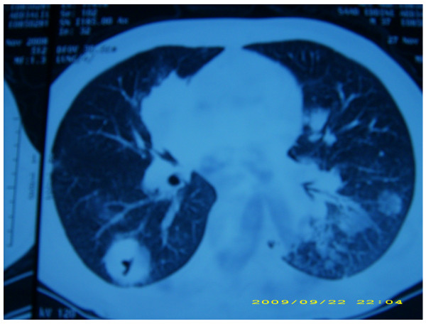
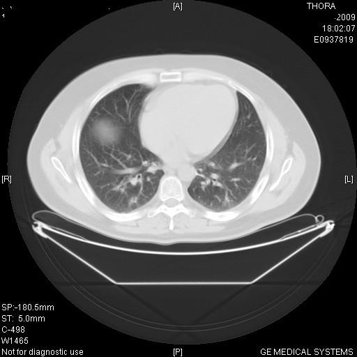
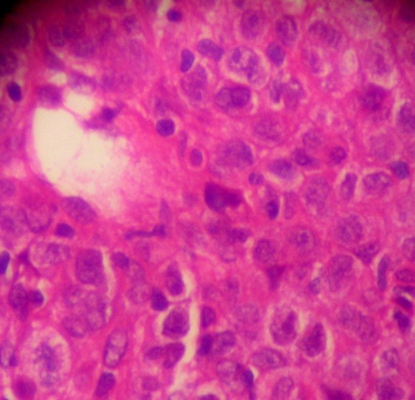
The first one is 39-year-old man in whom cholecystectomy made the diagnosis of primary non-Hodgkin's lymphoma of the gallbladder. He presented in chest CT scan excavated nodules that had been biopsied and confirmed the diagnosis of non hodgkin lymphoma. He underwent 8 courses of chemotherapy CHOP 21 with complete remission. The second one is an 21 years old man who presented a right leg osteoblastic osteosarcoma with only excavated pulmonary nodules in extension assessment. He had 3 courses of polychemotherapy API (doxorubicin, platinum, and ifosfamide) with partial response. Unfortunately, he died following a septic shock.Review of the literature shows that excavated pulmonary nodules as metastasis are rare but we should consider this diagnosis every time we are in front of a cancer. Chest computed tomography is the best diagnosis imaging that could make this diagnosis. Differential diagnosis between benign and malignant bullous lesions is important because surgical excision affects survival in some malignancies.

CONCLUSIONS

Although pulmonary nodules are the most common cancer metastasis, a differential diagnosis of a concurrent primary malignancy should always be considered every time we have excavated lesions, even in patients with known malignant disease. Thorough chest evaluation is important, as multiple primary malignancies may occur concomitantly.

摘要

背景

挖掘性肺部转移较为罕见。我们报告两例经证实为骨肉瘤和胆囊淋巴瘤转移的挖掘性肺部结节病例。

病例介绍

第一个病例是一名 39 岁男性,因胆囊切除术诊断为原发性非霍奇金淋巴瘤。他的胸部 CT 扫描显示有挖掘性结节,经活检证实为非霍奇金淋巴瘤。他接受了 8 个周期的 CHOP 21 化疗,完全缓解。第二个病例是一名 21 岁男性,右下肢成骨肉瘤,仅在扩展评估时发现挖掘性肺部结节。他接受了 3 个周期的多药化疗 API(多柔比星、铂和异环磷酰胺),部分缓解。不幸的是,他因感染性休克死亡。文献回顾表明,挖掘性肺部结节作为转移较为罕见,但每次面对癌症患者时,我们都应考虑这种诊断。胸部计算机断层扫描是做出这一诊断的最佳诊断影像学方法。良性和恶性大疱性病变之间的鉴别诊断很重要,因为在某些恶性肿瘤中,手术切除会影响生存。

结论

尽管肺部结节是最常见的癌症转移,但每次遇到挖掘性病变时,即使患者患有已知的恶性疾病,也应始终考虑同时存在原发性恶性肿瘤的鉴别诊断。彻底的胸部评估很重要,因为可能同时发生多种原发性恶性肿瘤。

https://cdn.ncbi.nlm.nih.gov/pmc/blobs/bdf3/2904264/4d265930afce/1471-2342-10-13-3.jpg
https://cdn.ncbi.nlm.nih.gov/pmc/blobs/bdf3/2904264/4bde85adc7ef/1471-2342-10-13-1.jpg
https://cdn.ncbi.nlm.nih.gov/pmc/blobs/bdf3/2904264/4f0743b3bedf/1471-2342-10-13-2.jpg
https://cdn.ncbi.nlm.nih.gov/pmc/blobs/bdf3/2904264/4d265930afce/1471-2342-10-13-3.jpg
https://cdn.ncbi.nlm.nih.gov/pmc/blobs/bdf3/2904264/4bde85adc7ef/1471-2342-10-13-1.jpg
https://cdn.ncbi.nlm.nih.gov/pmc/blobs/bdf3/2904264/4f0743b3bedf/1471-2342-10-13-2.jpg
https://cdn.ncbi.nlm.nih.gov/pmc/blobs/bdf3/2904264/4d265930afce/1471-2342-10-13-3.jpg

相似文献

1
Excavated pulmonary nodules: an unusual clinical presentation of lung metastasis in two cases.肺部结节的挖掘:两例肺转移瘤的不常见临床表现。
BMC Med Imaging. 2010 Jun 24;10:13. doi: 10.1186/1471-2342-10-13.
2
Unusual cause of recurrent pneumothorax: excavated metastasis of osteosarcoma.复发性气胸的罕见病因:骨肉瘤的空洞性转移
Ann Thorac Surg. 2001 Dec;72(6):2111-3. doi: 10.1016/s0003-4975(01)02694-7.
3
[Recurrent spontaneous pneumothorax complicating osteosarcoma pulmonary metastasis].[复发性自发性气胸并发骨肉瘤肺转移]
Rev Pneumol Clin. 2005 Feb;61(1 Pt 1):50-2. doi: 10.1016/s0761-8417(05)84784-4.
4
Unusual thoracic CT manifestations of osteosarcoma: review of 16 cases.骨肉瘤的不典型胸部CT表现:16例病例回顾
Pediatr Radiol. 2008 May;38(5):551-8. doi: 10.1007/s00247-007-0735-3. Epub 2008 Feb 2.
5
Minimally invasive thoracoscopic ultrasound for localization of pulmonary nodules in children.微创胸腔镜超声用于儿童肺结节的定位
J Pediatr Surg. 2008 Dec;43(12):2315-22. doi: 10.1016/j.jpedsurg.2008.08.031.
6
Pulmonary nodules in patients with esophageal carcinoma.食管癌患者的肺结节
J Clin Gastroenterol. 1998 Jun;26(4):245-8. doi: 10.1097/00004836-199806000-00004.
7
[An extraosseous osteoblastic osteosarcoma in the breast mimicking a pulmonary coin lesion on the chest radiograph (author's transl)].乳腺骨外成骨性骨肉瘤在胸部X线片上酷似肺内结节(作者译)
Radiologe. 1979 May;19(5):189-92.
8
[Case of osteosarcoma of the pulmonary artery].[肺动脉骨肉瘤病例]
Nihon Kokyuki Gakkai Zasshi. 2010 Dec;48(12):923-9.
9
Accuracy of 16-channel multi-detector row chest computed tomography with thin sections in the detection of metastatic pulmonary nodules.16排多探测器胸部薄层计算机断层扫描在检测肺转移瘤结节中的准确性
Eur J Cardiothorac Surg. 2008 Mar;33(3):473-9. doi: 10.1016/j.ejcts.2007.12.011. Epub 2008 Jan 25.
10
Evaluation and management of solitary and multiple pulmonary nodules.孤立性和多发性肺结节的评估与管理
Clin Chest Med. 1992 Mar;13(1):83-95.

本文引用的文献

1
Massive cavitation of solid pulmonary metastatic lesions in a breast cancer patient: a case report.
Ann Oncol. 2002 Jan;13(1):173-4. doi: 10.1093/annonc/mdf001.
2
[Multiple cavitary pulmonary metastases from ovarian cancer: a case report].[卵巢癌多发空洞性肺转移:一例报告]
Nihon Kokyuki Gakkai Zasshi. 2001 Jun;39(6):430-3.
3
Atypical pulmonary metastases: spectrum of radiologic findings.非典型肺转移瘤:影像学表现谱
Radiographics. 2001 Mar-Apr;21(2):403-17. doi: 10.1148/radiographics.21.2.g01mr17403.
4
Gallbladder carcinoma: radiologic-pathologic correlation.胆囊癌:放射学与病理学的相关性
Radiographics. 2001 Mar-Apr;21(2):295-314; questionnaire, 549-55. doi: 10.1148/radiographics.21.2.g01mr16295.
5
Multiple pulmonary metastases with cavitation from gallbladder cancer.胆囊癌伴空洞形成的多发性肺转移
Intern Med. 1998 Mar;37(3):292-4. doi: 10.2169/internalmedicine.37.292.