Martins Delio Eulalio, Oliveira Valdeci Manoel de, Alves Maria Teresa de Seixas, Wajchenberg Marcelo, Landim Elcio, Belloti João Carlos, Puertas Eduardo Barros, Ishida Akira
Department of Orthopedics and Traumatology, Universidade Federal de São Paulo, São Paulo, Brazil.
Sao Paulo Med J. 2010;128(2):63-8. doi: 10.1590/s1516-31802010000200004.
There is controversy regarding which imaging method is best for identifying early degenerative alterations in intervertebral discs. No correlations between such methods and histological finds are presented in the literature. The aim of this study was to correlate the thickness of intervertebral discs measured on simple radiographs with the degree of degeneration seen on magnetic resonance images and the histological findings relating to nerve ends inside the discs.
Cross-sectional correlation study on the lumbar spines of human cadavers, at Universidade Federal de São Paulo (Unifesp), São Paulo, Brazil.
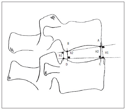
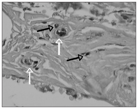
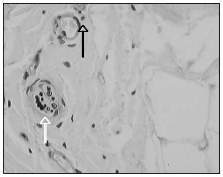
Ten lumbar spinal columns were extracted from human cadavers and subjected to magnetic resonance imaging and simple radiography. They were classified according to the degree of disc degeneration seen on magnetic resonance, and the thickness of the discs was measured on radiographs. The intervertebral discs were then extracted, embedded in paraffin and analyzed immunohistochemically with protein S100, and the nerve fibers were counted and classified.
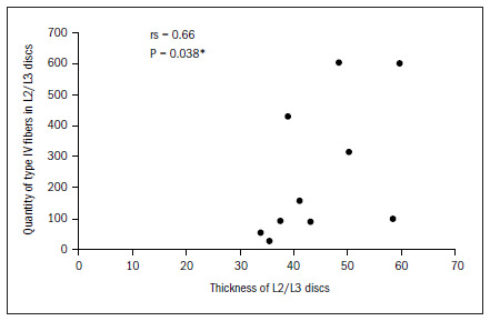
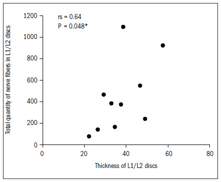
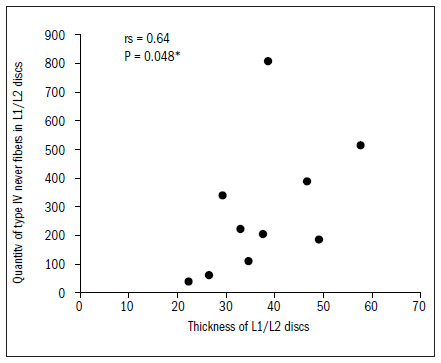
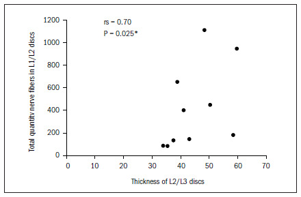
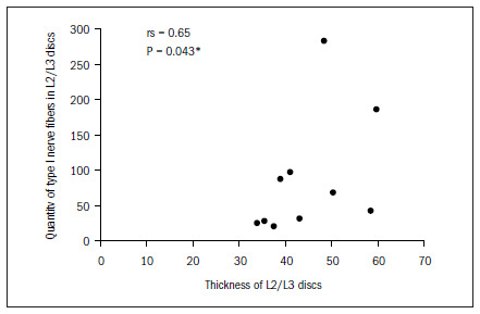
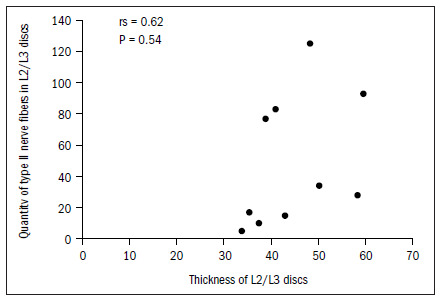
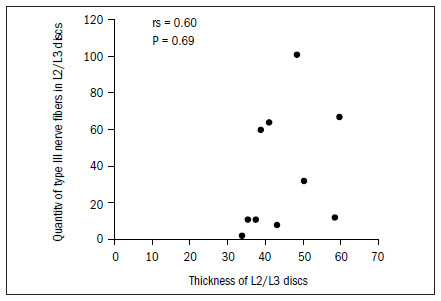
No correlation was observed between the thickness of the intervertebral discs and the degree of degeneration seen on magnetic resonance images. Only the uppermost lumbar discs (L1/L2 and L2/L3) presented a correlation between their thickness and type I and IV nerve endings.
Reduced disc thickness is unrelated to increased presence of nerve ends in intervertebral discs, or to the degree of disc degeneration.
关于哪种成像方法最适合识别椎间盘早期退变改变存在争议。文献中未提及此类方法与组织学发现之间的相关性。本研究的目的是将简单X线片上测量的椎间盘厚度与磁共振图像上显示的退变程度以及与椎间盘内神经末梢相关的组织学发现进行关联。
在巴西圣保罗的圣保罗联邦大学(Unifesp)对人类尸体腰椎进行横断面相关性研究。
从人类尸体中提取10个腰椎柱,进行磁共振成像和简单X线摄影。根据磁共振上显示的椎间盘退变程度进行分类,并在X线片上测量椎间盘厚度。然后取出椎间盘,石蜡包埋,用蛋白S100进行免疫组织化学分析,对神经纤维进行计数和分类。
未观察到椎间盘厚度与磁共振图像上显示的退变程度之间存在相关性。仅最上部的腰椎间盘(L1/L2和L2/L3)的厚度与I型和IV型神经末梢之间存在相关性。
椎间盘厚度减小与椎间盘内神经末梢数量增加或椎间盘退变程度无关。