Arizona Research Laboratories, University of Arizona, Tucson, Arizona, USA.
J Biomed Mater Res A. 2010 Dec 1;95(3):811-8. doi: 10.1002/jbm.a.32925.
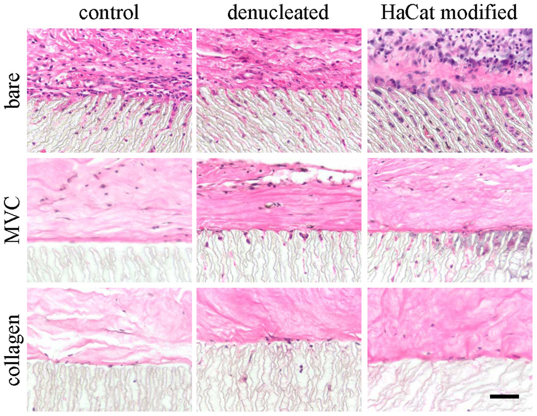
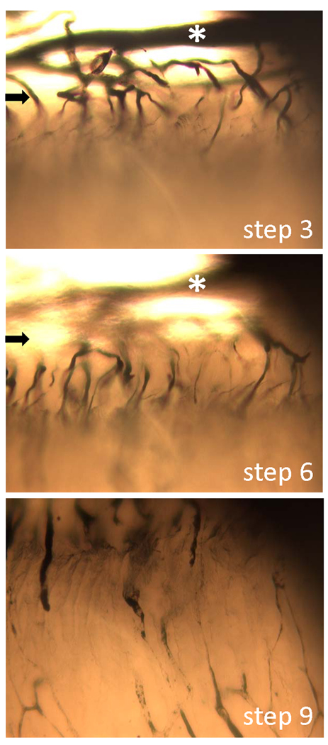
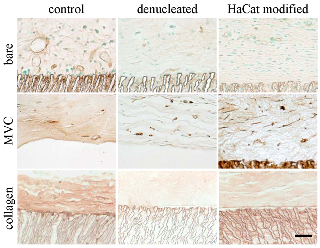
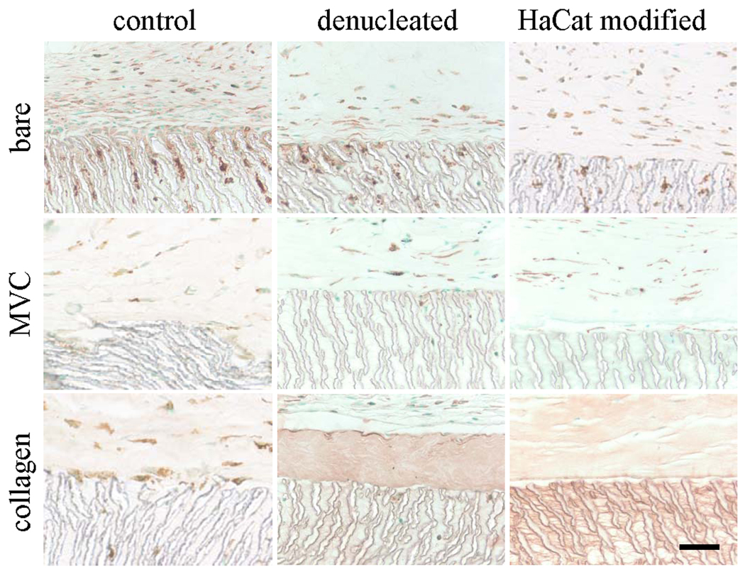
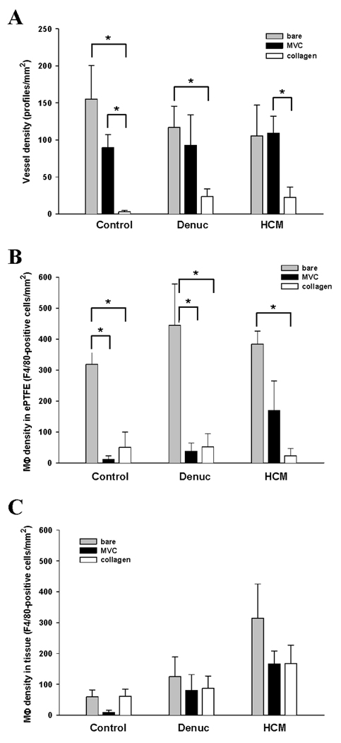
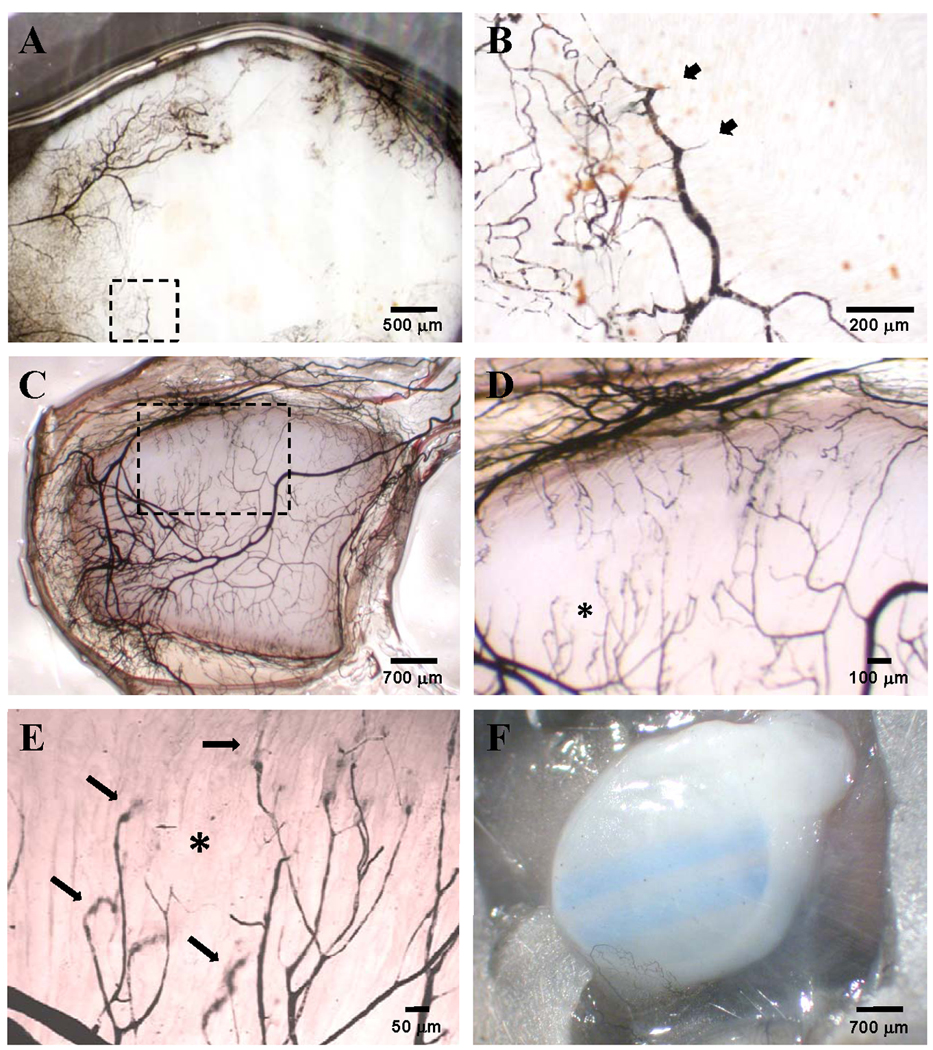
During the typical healing response to an implanted biomaterial, vascular-rich granulation tissue forms around the implant and later resolves into a relatively avascular, fibrous capsule. We have previously shown that a microvascular construct (MVC) consisting of isolated microvessel fragments suspended in a collagen I gel forms a persistent microcirculation in lieu of avascular scar when implanted. The current study evaluated the potential for microvascular constructs to maintain a vascularized tissue environment around an implanted biomaterial. An analysis of the peri-implant tissue around bare expanded polytetrafluoroethylene (ePTFE), ePTFE embedded within a microvascular construct, or ePTFE embedded within collagen alone revealed that the presence of the MVC, but not collagen alone, promoted vascular densities comparable to that of the granulation tissue formed around bare ePTFE. The vessels within the microvascular construct surrounding the ePTFE were perfusion competent, as determined by India ink perfusion casting, and extended into the interstices of the polymer. In contrast to bare ePTFE, the presence of the MVC or collagen alone significantly reduced the number of activated macrophages in association with ePTFE. Similar results were observed for ePTFE modified to increase cellularity and prevent the formation of an avascular scar. The microvascular construct may prove effective in forming vascularized tissue environments and limiting the number of activated macrophages around implanted polymers thereby leading to effective implant incorporation.
在植入生物材料后的典型愈合反应过程中,富含血管的肉芽组织围绕植入物形成,随后逐渐转化为相对无血管的纤维囊。我们之前已经证明,由悬浮在 I 型胶原凝胶中的分离微血管片段组成的微血管构建体(MVC)在植入时会形成持久的微循环,而不是无血管的瘢痕。本研究评估了微血管构建体在植入生物材料周围维持血管化组织环境的潜力。对裸露的膨体聚四氟乙烯(ePTFE)、嵌入微血管构建体中的 ePTFE 或仅嵌入胶原中的 ePTFE 周围的植入物组织进行分析后发现,MVC 的存在,而不是单独的胶原,促进了血管密度与裸露的 ePTFE 周围形成的肉芽组织相当。通过印度墨水灌注铸型法确定,围绕 ePTFE 的 MVC 中的血管具有灌注能力,并延伸到聚合物的间隙中。与裸露的 ePTFE 相比,MVC 或单独的胶原的存在显著减少了与 ePTFE 相关的活化巨噬细胞的数量。对增加细胞密度并防止无血管瘢痕形成的 ePTFE 进行改性时,也观察到了类似的结果。微血管构建体可能在形成血管化组织环境和限制植入聚合物周围活化巨噬细胞的数量方面有效,从而导致有效的植入物整合。