Orthodontics, Federal University of Ceará, Fortaleza, CE, Brazil.
J Appl Oral Sci. 2010 Jul-Aug;18(4):335-42. doi: 10.1590/s1678-77572010000400003.
The aim of this study was to test the efficacy of a locally applied 8.5% nanostructured doxycycline (DOX) gel in preventing alveolar bone loss in experimental periodontal disease (EPD) in rats by using the tapping mode atomic force microscopy (AFM).
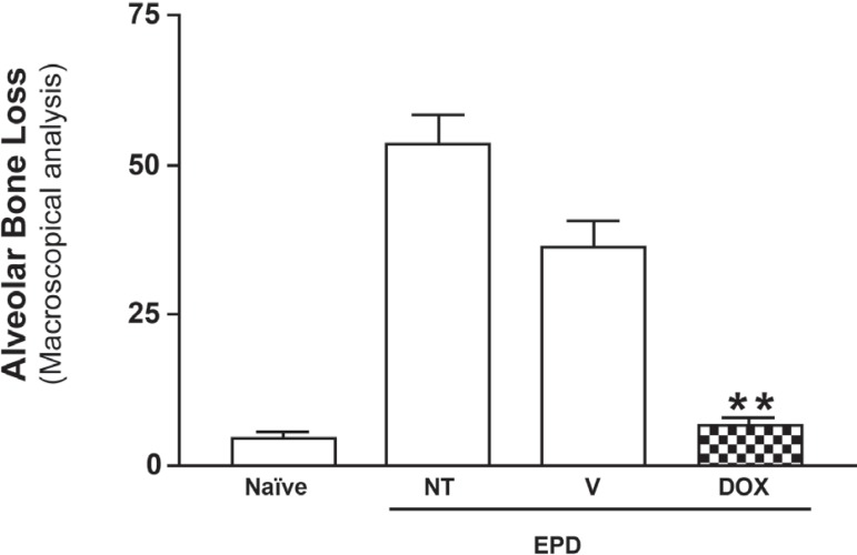
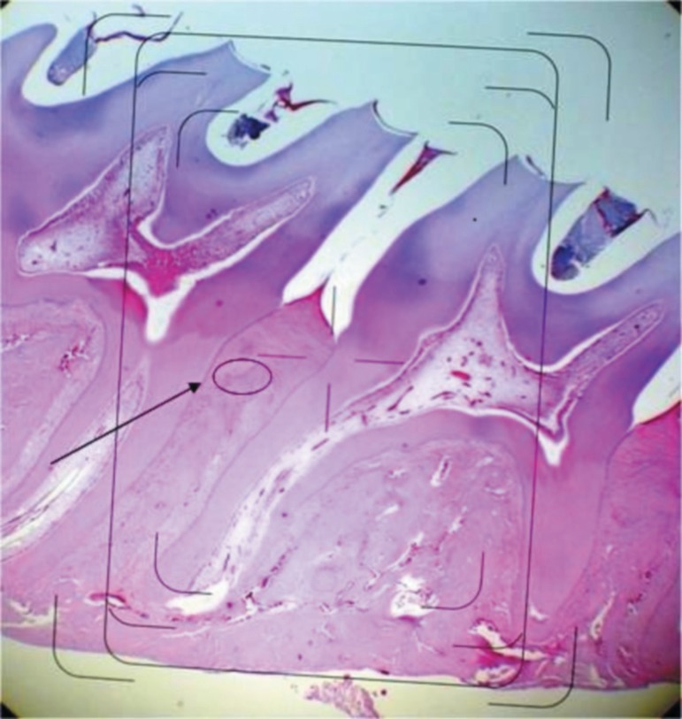
EPD was induced in 24 Wistar rats. Animals were treated with the doxycycline gel topically, immediately after EPD induction, and 3 times a day during 11 days. Four groups (n=6) were formed as follows: Naïve group (animals not subjected to EPD nor treated); non-treated (NT) group (animals subjected to EPD, but not treated); vehicle gel (VG) group (animals subjected to EPD and treated with topical gel vehicle); and DOX group (test group): animals subjected to EPD and treated with the 8.5% DOX gel. In order to investigate topographical changes in histological sections, a novel simple method was used for sample preparation, by etching sections from paraffin-embedded specimens with xylol.
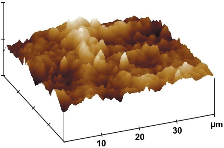
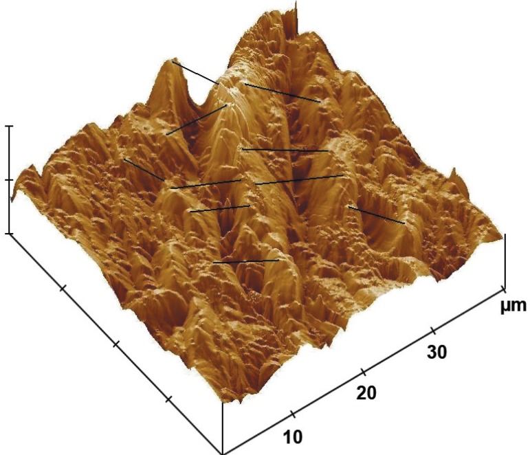
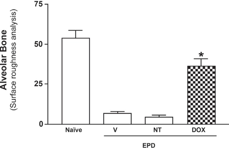
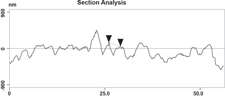
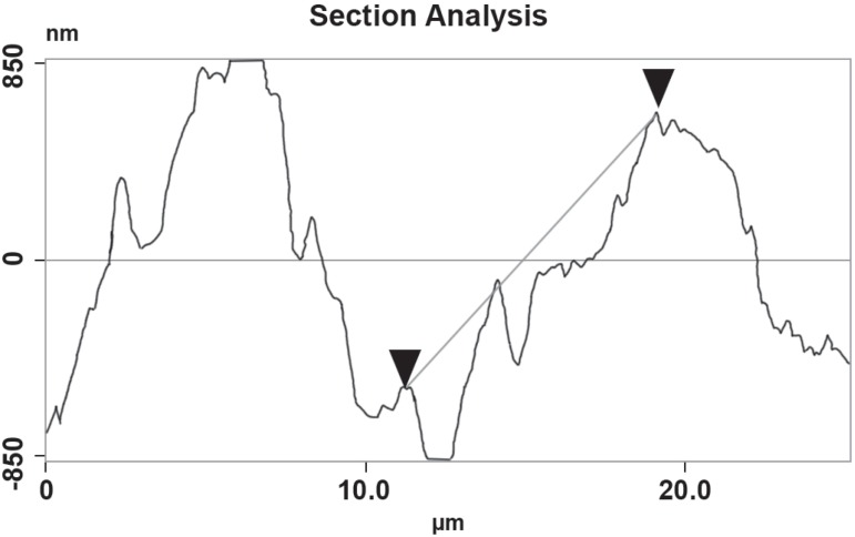
Comparing the AFM images, several grooves were observed on the surface of the alveolar bone and other periodontal structures in the NT and VG groups, with significantly greater depths when compared to the DOX group (p<0.05).
Periodontal structures were brought into high relief confirming to be a simple and cost-effective method for AFM imaging with ultrastructural resolution. The doxycycline gel was able to afford periodontal surface preservation, with flatter grooves.
本研究旨在通过敲击模式原子力显微镜(AFM)测试局部应用 8.5%纳米结构多西环素(DOX)凝胶在预防实验性牙周病(EPD)大鼠牙槽骨丢失中的疗效。
在 24 只 Wistar 大鼠中诱导 EPD。动物在 EPD 诱导后立即局部给予 DOX 凝胶治疗,并在 11 天内每天治疗 3 次。形成以下 4 组(n=6):未处理组(未接受 EPD 治疗的动物);未治疗组(接受 EPD 治疗但未治疗的动物);载体凝胶组(接受 EPD 治疗并用局部凝胶载体治疗的动物);和 DOX 组(实验组):接受 EPD 治疗并用 8.5% DOX 凝胶治疗的动物。为了研究组织学切片的形貌变化,采用了一种新颖的简单方法进行样本制备,即用二甲苯蚀刻石蜡包埋标本的切片。
通过 AFM 图像比较,在 NT 和 VG 组中,牙槽骨和其他牙周结构的表面观察到几个凹槽,与 DOX 组相比,深度明显更大(p<0.05)。
牙周结构被突出显示出来,证实这是一种具有超微结构分辨率的简单且具有成本效益的 AFM 成像方法。多西环素凝胶能够保持牙周表面的完整性,凹槽更平坦。