• 文献检索
  • 文档翻译
  • 深度研究
  • 学术资讯
  • Suppr Zotero 插件Zotero 插件
  • 邀请有礼
  • 套餐&价格
  • 历史记录
应用&插件
Suppr Zotero 插件Zotero 插件浏览器插件Mac 客户端Windows 客户端微信小程序
定价
高级版会员购买积分包购买API积分包
服务
文献检索文档翻译深度研究API 文档MCP 服务
关于我们
关于 Suppr公司介绍联系我们用户协议隐私条款
关注我们

Suppr 超能文献

核心技术专利:CN118964589B侵权必究
粤ICP备2023148730 号-1Suppr @ 2026

文献检索

告别复杂PubMed语法,用中文像聊天一样搜索,搜遍4000万医学文献。AI智能推荐,让科研检索更轻松。

立即免费搜索

文件翻译

保留排版,准确专业,支持PDF/Word/PPT等文件格式,支持 12+语言互译。

免费翻译文档

深度研究

AI帮你快速写综述,25分钟生成高质量综述,智能提取关键信息,辅助科研写作。

立即免费体验

参考稳态自由进动心血管磁共振测量左心房的尺寸和容积。

Reference left atrial dimensions and volumes by steady state free precession cardiovascular magnetic resonance.

机构信息

Cardiac Imaging Unit, ERESA, Valencia, Spain.

出版信息

J Cardiovasc Magn Reson. 2010 Nov 11;12(1):65. doi: 10.1186/1532-429X-12-65.

DOI:10.1186/1532-429X-12-65
PMID:21070636
原文链接:https://pmc.ncbi.nlm.nih.gov/articles/PMC2994941/
Abstract

BACKGROUND

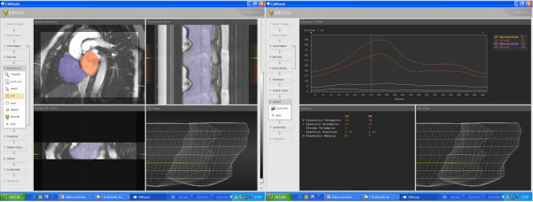
Left atrial (LA) size is related to cardiovascular morbidity and mortality. Cardiovascular magnetic resonance (CMR) provides high quality images of the left atrium with high temporal resolution steady state free precession (SSFP) cine sequences. We used SSFP cines to define normal ranges for LA volumes and dimensions relative to gender, age and body surface area (BSA), and examine the relative value of 2D atrial imaging techniques in patients.For definition of normal ranges of LA volume we studied 120 healthy subjects after careful exclusion of cardiovascular abnormality (60 men, 60 women; 20 subjects per age decile from 20 to 80 years). Data were generated from 3-dimensional modeling, including tracking of the atrioventricular ring motion and time-volume curves analysis. For definition of the best 2D images-derived predictors of LA enlargement, we studied 120 patients (60 men, 60 women; age range 20 to 80 years) with a clinical indication for CMR.

RESULTS

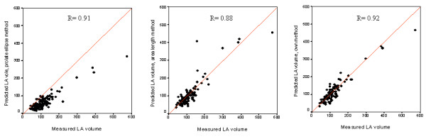
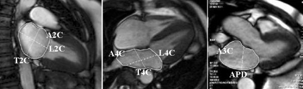
In the healthy subjects, age was associated with LA 4-chamber transverse and 3-chamber anteroposterior diameters, but not with LA volume. Gender was an independent predictor of most absolute LA dimensions and volume, but following normalization to BSA, some associations became non-significant. CMR normal ranges were modeled and are tabled for clinical use with normalization, where appropriate, for BSA and gender and display of parameter variation with age. The best 2D predictors of LA volume were the 2-chamber area and 3-chamber area (both r = 0.90, p < 0.001).

CONCLUSIONS

These CMR data show that LA dimensions and volume in healthy, individuals vary significantly by BSA, with lesser effects of age and gender.

摘要

背景

左心房(LA)的大小与心血管发病率和死亡率有关。心血管磁共振(CMR)提供了具有高时间分辨率稳态自由进动(SSFP)电影序列的高质量左心房图像。我们使用 SSFP 电影来定义与性别、年龄和体表面积(BSA)相关的 LA 容积和尺寸的正常范围,并检查 2D 心房成像技术在患者中的相对价值。为了定义 LA 容积的正常范围,我们在仔细排除心血管异常(60 名男性,60 名女性;每个年龄十分位数有 20 名受试者,年龄范围为 20 至 80 岁)后研究了 120 名健康受试者。数据来自三维建模,包括房室环运动的跟踪和时容积曲线分析。为了定义最佳的 2D 图像衍生的 LA 扩大预测因子,我们研究了 120 名有 CMR 临床指征的患者(60 名男性,60 名女性;年龄范围为 20 至 80 岁)。

结果

在健康受试者中,年龄与 LA 四腔横径和三腔前后径相关,但与 LA 容积无关。性别是大多数绝对 LA 尺寸和容积的独立预测因子,但在归一化到 BSA 后,一些关联变得不显著。CMR 正常范围进行了建模,并以表格形式展示,以便临床使用,包括适当的 BSA 和性别归一化以及参数随年龄的变化显示。LA 容积的最佳 2D 预测因子是两腔面积和三腔面积(两者 r = 0.90,p < 0.001)。

结论

这些 CMR 数据表明,在健康个体中,LA 的尺寸和容积因 BSA 而有显著差异,而年龄和性别影响较小。

https://cdn.ncbi.nlm.nih.gov/pmc/blobs/91b3/2994941/dcee39846303/1532-429X-12-65-3.jpg
https://cdn.ncbi.nlm.nih.gov/pmc/blobs/91b3/2994941/e126bdbaa668/1532-429X-12-65-1.jpg
https://cdn.ncbi.nlm.nih.gov/pmc/blobs/91b3/2994941/d3cf0dcee9df/1532-429X-12-65-2.jpg
https://cdn.ncbi.nlm.nih.gov/pmc/blobs/91b3/2994941/dcee39846303/1532-429X-12-65-3.jpg
https://cdn.ncbi.nlm.nih.gov/pmc/blobs/91b3/2994941/e126bdbaa668/1532-429X-12-65-1.jpg
https://cdn.ncbi.nlm.nih.gov/pmc/blobs/91b3/2994941/d3cf0dcee9df/1532-429X-12-65-2.jpg
https://cdn.ncbi.nlm.nih.gov/pmc/blobs/91b3/2994941/dcee39846303/1532-429X-12-65-3.jpg

相似文献

1
Reference left atrial dimensions and volumes by steady state free precession cardiovascular magnetic resonance.参考稳态自由进动心血管磁共振测量左心房的尺寸和容积。
J Cardiovasc Magn Reson. 2010 Nov 11;12(1):65. doi: 10.1186/1532-429X-12-65.
2
Reference right atrial dimensions and volume estimation by steady state free precession cardiovascular magnetic resonance.稳态自由进动心血管磁共振右心房大小和容量评估的参考。
J Cardiovasc Magn Reson. 2013 Apr 8;15(1):29. doi: 10.1186/1532-429X-15-29.
3
Characterization of left and right atrial function in healthy volunteers by cardiovascular magnetic resonance.通过心血管磁共振对健康志愿者左、右心房功能的特征描述。
J Cardiovasc Magn Reson. 2016 Oct 10;18(1):64. doi: 10.1186/s12968-016-0284-8.
4
Quantification of the left atrium applying cardiovascular magnetic resonance in clinical routine.在临床常规中应用心血管磁共振对左心房进行定量分析。
Scand Cardiovasc J. 2018 Apr;52(2):85-92. doi: 10.1080/14017431.2017.1423107. Epub 2018 Jan 5.
5
Determination of normal gender-specific left atrial dimensions by cardiovascular magnetic resonance imaging.通过心血管磁共振成像确定正常的性别特异性左心房尺寸。
J Cardiovasc Magn Reson. 2005;7(4):677-83. doi: 10.1081/jcmr-65621.
6
Left atrial volume index in healthy subjects: clinical and echocardiographic correlates.健康受试者的左心房容积指数:临床及超声心动图相关性
Echocardiography. 2013 Oct;30(9):1001-7. doi: 10.1111/echo.12217. Epub 2013 Apr 18.
7
Quantification of left atrial volumes using three-dimensional wall motion tracking echocardiographic technology: comparison with cardiac magnetic resonance.使用三维心动图壁运动跟踪技术定量左心房容积:与心脏磁共振的比较。
Eur Heart J Cardiovasc Imaging. 2014 Jul;15(7):793-9. doi: 10.1093/ehjci/jeu001. Epub 2014 Jan 30.
8
Left Atrial Structure in Relationship to Age, Sex, Ethnicity, and Cardiovascular Risk Factors: MESA (Multi-Ethnic Study of Atherosclerosis).与年龄、性别、种族和心血管危险因素相关的左心房结构:动脉粥样硬化多民族研究(MESA)
Circ Cardiovasc Imaging. 2017 Feb;10(2). doi: 10.1161/CIRCIMAGING.116.005379.
9
Quantification of left atrial strain and strain rate using Cardiovascular Magnetic Resonance myocardial feature tracking: a feasibility study.使用心血管磁共振心肌特征追踪技术对左心房应变和应变率进行定量分析:一项可行性研究。
J Cardiovasc Magn Reson. 2014 Aug 12;16(1):60. doi: 10.1186/s12968-014-0060-6.
10
Reference value of left and right atrial size and phasic function by SSFP CMR at 3.0 T in healthy Chinese adults.健康中国成年人 3.0T 下稳态自由进动 CMR 测量左、右心房大小和时相功能的参考值。
Sci Rep. 2017 Jun 9;7(1):3196. doi: 10.1038/s41598-017-03377-6.

引用本文的文献

1
Atrial fibrillation and thromboembolic risk in hypertrophic cardiomyopathy.肥厚型心肌病中的心房颤动与血栓栓塞风险
J Cardiovasc Imaging. 2025 Aug 25;33(1):12. doi: 10.1186/s44348-025-00057-2.
2
Is regional atrial strain a useful surrogate of regional atrial fibrosis in atrial cardiomyopathy?在心房心肌病中,局部心房应变是否是局部心房纤维化的有用替代指标?
Eur Heart J Imaging Methods Pract. 2025 May 28;3(2):qyaf068. doi: 10.1093/ehjimp/qyaf068. eCollection 2025 Jul.
3
Enhanced risk stratification in chronic coronary syndrome using the left atrioventricular coupling index.

本文引用的文献

1
Cardiac chamber volumes, function, and mass as determined by 64-multidetector row computed tomography: mean values among healthy adults free of hypertension and obesity.64排多层螺旋计算机断层扫描测定的心脏腔室容积、功能及质量:无高血压和肥胖的健康成年人的平均值
JACC Cardiovasc Imaging. 2008 Nov;1(6):782-6. doi: 10.1016/j.jcmg.2008.04.015.
2
Left atrial volume measurement with automated border detection by 3-dimensional echocardiography: comparison with Magnetic Resonance Imaging.三维超声心动图自动边界检测法测量左心房容积:与磁共振成像的比较
Cardiovasc Ultrasound. 2009 Mar 31;7:16. doi: 10.1186/1476-7120-7-16.
3
使用左房室耦合指数对慢性冠状动脉综合征进行强化风险分层。
Clin Res Cardiol. 2025 Jun 23. doi: 10.1007/s00392-025-02699-0.
4
Early cardiac involvement in patients with hyperthyroidism evaluated by cardiac magnetic resonance feature tracking.通过心脏磁共振特征追踪评估甲状腺功能亢进症患者的早期心脏受累情况。
Front Endocrinol (Lausanne). 2025 Jun 2;16:1566029. doi: 10.3389/fendo.2025.1566029. eCollection 2025.
5
The role of left atrio-ventricular coupling index and left atrial ejection fraction in predicting onset of atrial fibrillation and adverse cardiac events in hypertrophic cardiomyopathy.左房室耦合指数和左心房射血分数在预测肥厚型心肌病患者房颤发作及不良心脏事件中的作用
Cardiovasc Ultrasound. 2025 Mar 28;23(1):10. doi: 10.1186/s12947-025-00343-5.
6
Comprehensive reference ranges for cardiovascular magnetic resonance: time to move on from single centre data?心血管磁共振的综合参考范围:是时候摒弃单中心数据了吗?
Int J Cardiovasc Imaging. 2025 May;41(5):913-932. doi: 10.1007/s10554-025-03370-5. Epub 2025 Mar 27.
7
Correlating Left Atrial Enlargement and Left Ventricular Hypertrophy on ECG With Echocardiography and Cardiac Magnetic Resonance Imaging.心电图上左心房扩大和左心室肥厚与超声心动图及心脏磁共振成像的相关性
J Am Heart Assoc. 2025 Mar 18;14(6):e037772. doi: 10.1161/JAHA.124.037772. Epub 2025 Mar 13.
8
Society for Cardiovascular Magnetic Resonance reference values ("normal values") in cardiovascular magnetic resonance: 2025 update.心血管磁共振学会心血管磁共振参考值(“正常值”):2025年更新。
J Cardiovasc Magn Reson. 2025;27(1):101853. doi: 10.1016/j.jocmr.2025.101853. Epub 2025 Feb 4.
9
A novel network with enhanced edge information for left atrium segmentation from LGE-MRI.一种用于从LGE-MRI中进行左心房分割的具有增强边缘信息的新型网络。
Front Physiol. 2024 Dec 10;15:1478347. doi: 10.3389/fphys.2024.1478347. eCollection 2024.
10
Cardiac MRI in heart failure with preserved ejection fraction.心脏磁共振成像在射血分数保留型心力衰竭中的应用。
Radiol Med. 2024 Oct;129(10):1468-1484. doi: 10.1007/s11547-024-01874-z. Epub 2024 Aug 19.
Quantitative assessment of left atrial volume by electrocardiographic-gated contrast-enhanced multidetector computed tomography.
通过心电图门控对比增强多层螺旋计算机断层扫描对左心房容积进行定量评估。
J Cardiovasc Comput Tomogr. 2009 Mar-Apr;3(2):80-7. doi: 10.1016/j.jcct.2009.02.002. Epub 2009 Feb 14.
4
Relation of left atrial volume from three-dimensional computed tomography to atrial fibrillation recurrence following ablation.三维计算机断层扫描测量的左心房容积与消融术后房颤复发的关系。
Am J Cardiol. 2009 Apr 1;103(7):989-93. doi: 10.1016/j.amjcard.2008.12.021.
5
Left atrial remodelling in patients with myocardial infarction complicated by heart failure, left ventricular dysfunction, or both: the VALIANT Echo study.心肌梗死合并心力衰竭、左心室功能障碍或两者兼具患者的左心房重塑:VALIANT回声研究
Eur Heart J. 2009 Jan;30(1):56-65. doi: 10.1093/eurheartj/ehn499. Epub 2008 Nov 11.
6
How many patients would be misclassified using M-mode and two-dimensional estimates of left atrial size instead of left atrial volume? A three-dimensional echocardiographic study.与使用左心房容积相比,使用M型和二维测量左心房大小会导致多少患者被误诊?一项三维超声心动图研究。
J Cardiovasc Med (Hagerstown). 2008 May;9(5):476-84. doi: 10.2459/JCM.0b013e3282f194f0.
7
Effects of aging on left atrioventricular coupling and left ventricular filling assessed using cardiac magnetic resonance imaging in healthy subjects.在健康受试者中使用心脏磁共振成像评估衰老对左房室耦合和左心室充盈的影响。
Am J Cardiol. 2007 Jul 1;100(1):122-7. doi: 10.1016/j.amjcard.2007.02.060. Epub 2007 May 21.
8
Cardiovascular features of heart failure with preserved ejection fraction versus nonfailing hypertensive left ventricular hypertrophy in the urban Baltimore community: the role of atrial remodeling/dysfunction.巴尔的摩市区社区射血分数保留的心力衰竭与非衰竭高血压左心室肥厚的心血管特征:心房重塑/功能障碍的作用
J Am Coll Cardiol. 2007 Jan 16;49(2):198-207. doi: 10.1016/j.jacc.2006.08.050. Epub 2006 Dec 29.
9
Left atrial size and risk of major cardiovascular events during antihypertensive treatment: losartan intervention for endpoint reduction in hypertension trial.降压治疗期间左心房大小与主要心血管事件风险:氯沙坦干预降低高血压终点事件试验
Hypertension. 2007 Feb;49(2):311-6. doi: 10.1161/01.HYP.0000254322.96189.85. Epub 2006 Dec 18.
10
Reference right ventricular systolic and diastolic function normalized to age, gender and body surface area from steady-state free precession cardiovascular magnetic resonance.来自稳态自由进动心血管磁共振的、根据年龄、性别和体表面积进行标准化的右心室收缩和舒张功能参考值。
Eur Heart J. 2006 Dec;27(23):2879-88. doi: 10.1093/eurheartj/ehl336. Epub 2006 Nov 6.