• 文献检索
  • 文档翻译
  • 深度研究
  • 学术资讯
  • Suppr Zotero 插件Zotero 插件
  • 邀请有礼
  • 套餐&价格
  • 历史记录
应用&插件
Suppr Zotero 插件Zotero 插件浏览器插件Mac 客户端Windows 客户端微信小程序
定价
高级版会员购买积分包购买API积分包
服务
文献检索文档翻译深度研究API 文档MCP 服务
关于我们
关于 Suppr公司介绍联系我们用户协议隐私条款
关注我们

Suppr 超能文献

核心技术专利:CN118964589B侵权必究
粤ICP备2023148730 号-1Suppr @ 2026

文献检索

告别复杂PubMed语法,用中文像聊天一样搜索,搜遍4000万医学文献。AI智能推荐,让科研检索更轻松。

立即免费搜索

文件翻译

保留排版,准确专业,支持PDF/Word/PPT等文件格式,支持 12+语言互译。

免费翻译文档

深度研究

AI帮你快速写综述,25分钟生成高质量综述,智能提取关键信息,辅助科研写作。

立即免费体验

利用磁共振成像对肝脏脂肪进行定量分析。

Quantification of liver fat with magnetic resonance imaging.

作者信息

Reeder Scott B, Sirlin Claude B

机构信息

Liver Imaging Research Program, Department of Radiology, University of Wisconsin, E1/374 CSC, 600 Highland Avenue, Madison, WI 53792-3252, USA.

出版信息

Magn Reson Imaging Clin N Am. 2010 Aug;18(3):337-57, ix. doi: 10.1016/j.mric.2010.08.013.

DOI:10.1016/j.mric.2010.08.013
PMID:21094444
原文链接:https://pmc.ncbi.nlm.nih.gov/articles/PMC3002753/
Abstract

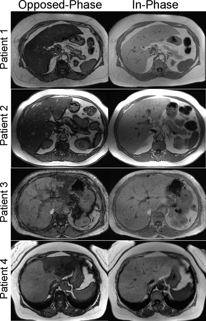
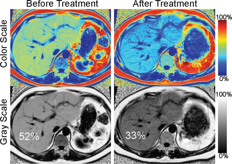
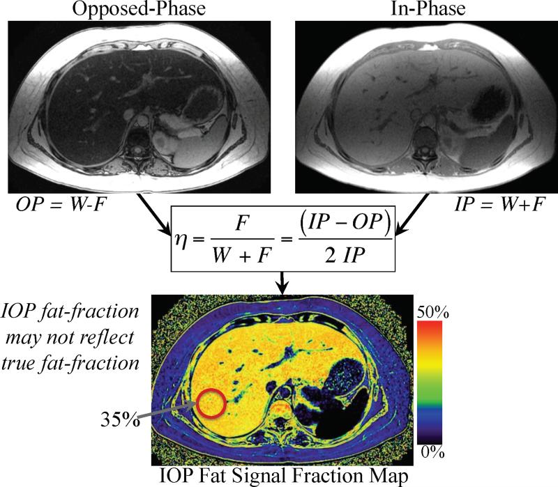
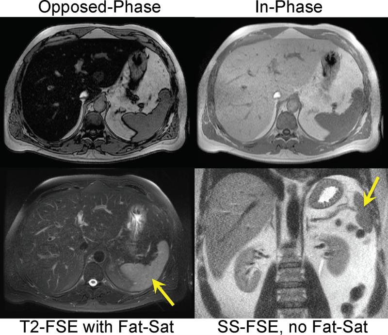
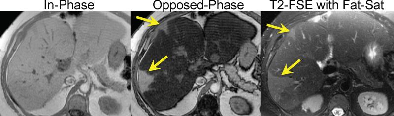
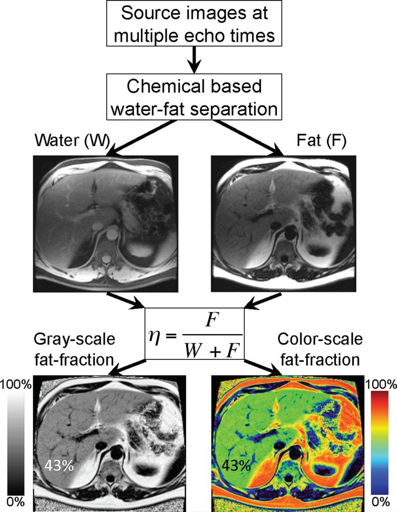
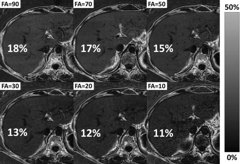
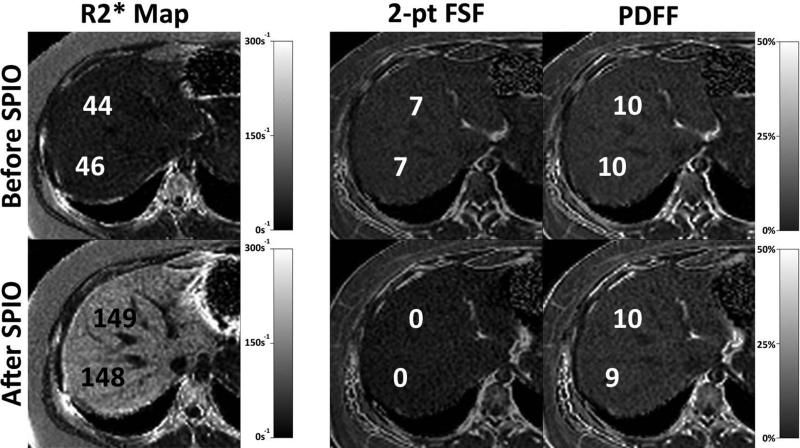
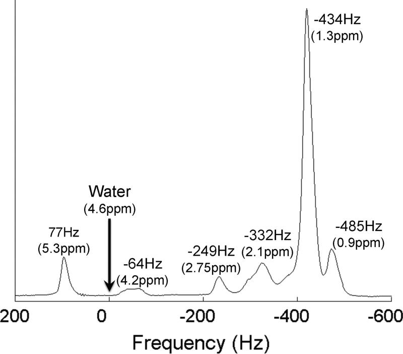
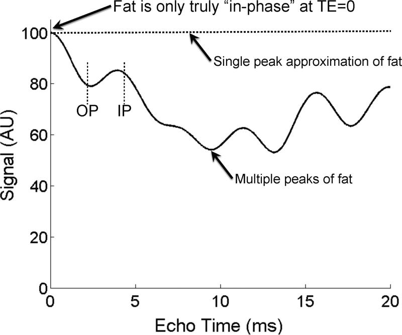
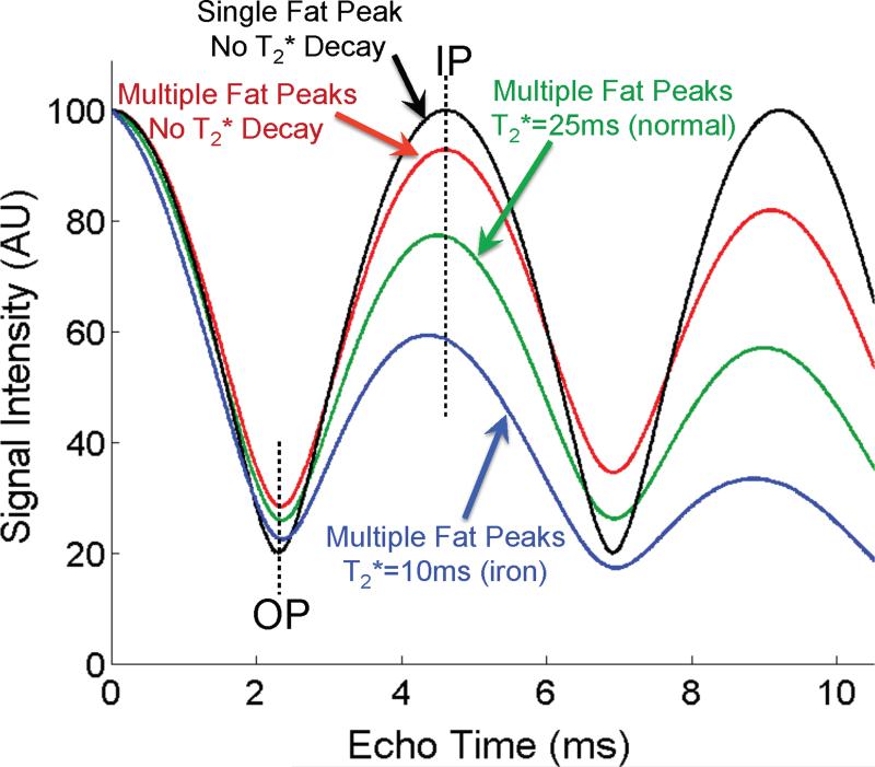
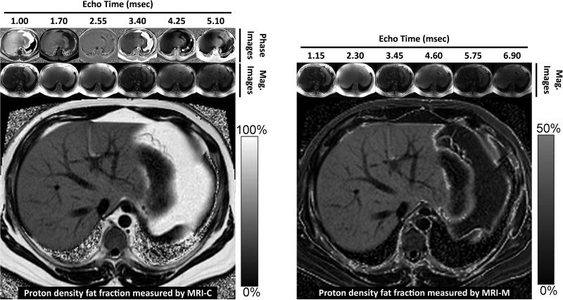
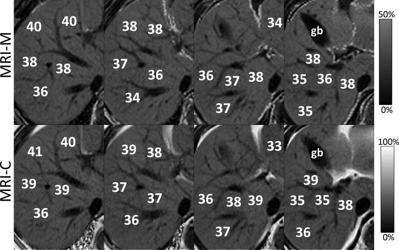
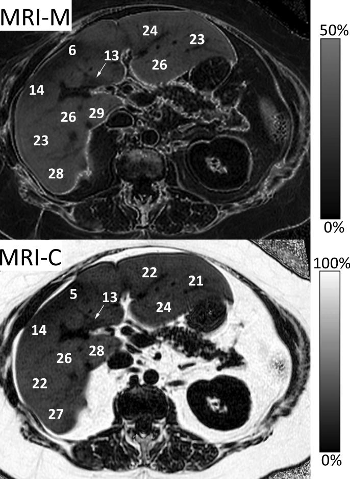
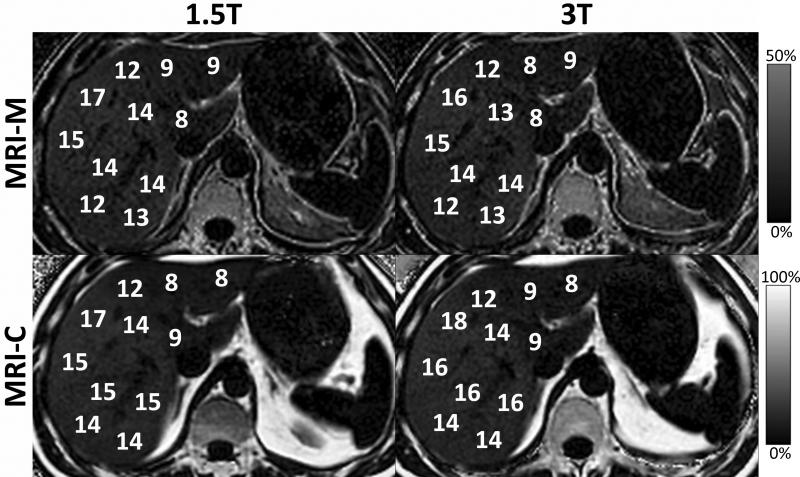
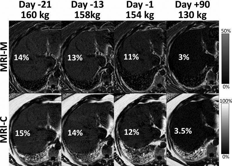
Intracellular fat accumulation is common feature of liver disease. Intracellular fat (steatosis) is the histologic hallmark of nonalcoholic fatty liver disease but also may occur with alcohol abuse, viral hepatitis, HIV and genetic lipodystrophies, and chemotherapy. This article reviews emerging MR imaging techniques that attempt to quantify liver fat. The content provides an overview of fatty liver disease and diseases where fat is an important disease feature. Also discussed is the current use and limitation of nontargeted biopsy in diffuse liver disease and why quantitative noninvasive biomarkers of liver fat would be beneficial.

摘要

细胞内脂肪堆积是肝脏疾病的常见特征。细胞内脂肪(脂肪变性)是非酒精性脂肪性肝病的组织学标志,但也可能发生于酒精滥用、病毒性肝炎、HIV感染、遗传性脂肪营养不良以及化疗过程中。本文综述了旨在定量肝脏脂肪的新兴磁共振成像技术。内容概述了脂肪性肝病以及脂肪作为重要疾病特征的疾病。还讨论了非靶向活检在弥漫性肝病中的当前应用及局限性,以及为什么肝脏脂肪的定量非侵入性生物标志物会有益处。

https://cdn.ncbi.nlm.nih.gov/pmc/blobs/ccb6/3002753/eab81562cd81/nihms-250182-f0016.jpg
https://cdn.ncbi.nlm.nih.gov/pmc/blobs/ccb6/3002753/62a79f878994/nihms-250182-f0001.jpg
https://cdn.ncbi.nlm.nih.gov/pmc/blobs/ccb6/3002753/14ee95eea3ba/nihms-250182-f0002.jpg
https://cdn.ncbi.nlm.nih.gov/pmc/blobs/ccb6/3002753/bbebf317542f/nihms-250182-f0003.jpg
https://cdn.ncbi.nlm.nih.gov/pmc/blobs/ccb6/3002753/8683e88b6fe1/nihms-250182-f0004.jpg
https://cdn.ncbi.nlm.nih.gov/pmc/blobs/ccb6/3002753/33d315306d25/nihms-250182-f0005.jpg
https://cdn.ncbi.nlm.nih.gov/pmc/blobs/ccb6/3002753/78bb7b4aaa28/nihms-250182-f0006.jpg
https://cdn.ncbi.nlm.nih.gov/pmc/blobs/ccb6/3002753/6bffb08eaf52/nihms-250182-f0007.jpg
https://cdn.ncbi.nlm.nih.gov/pmc/blobs/ccb6/3002753/18cf6ed61dae/nihms-250182-f0008.jpg
https://cdn.ncbi.nlm.nih.gov/pmc/blobs/ccb6/3002753/e5f683838845/nihms-250182-f0009.jpg
https://cdn.ncbi.nlm.nih.gov/pmc/blobs/ccb6/3002753/1e85c7a00e7e/nihms-250182-f0010.jpg
https://cdn.ncbi.nlm.nih.gov/pmc/blobs/ccb6/3002753/51bb91603aa5/nihms-250182-f0011.jpg
https://cdn.ncbi.nlm.nih.gov/pmc/blobs/ccb6/3002753/a6c513a78fcf/nihms-250182-f0012.jpg
https://cdn.ncbi.nlm.nih.gov/pmc/blobs/ccb6/3002753/099dae6270b1/nihms-250182-f0013.jpg
https://cdn.ncbi.nlm.nih.gov/pmc/blobs/ccb6/3002753/5672d960534e/nihms-250182-f0014.jpg
https://cdn.ncbi.nlm.nih.gov/pmc/blobs/ccb6/3002753/08657023f874/nihms-250182-f0015.jpg
https://cdn.ncbi.nlm.nih.gov/pmc/blobs/ccb6/3002753/eab81562cd81/nihms-250182-f0016.jpg

相似文献

1
Quantification of liver fat with magnetic resonance imaging.利用磁共振成像对肝脏脂肪进行定量分析。
Magn Reson Imaging Clin N Am. 2010 Aug;18(3):337-57, ix. doi: 10.1016/j.mric.2010.08.013.
2
Fatty liver disease: MR imaging techniques for the detection and quantification of liver steatosis.脂肪肝疾病:用于检测和量化肝脏脂肪变性的磁共振成像技术
Radiographics. 2009 Jan-Feb;29(1):231-60. doi: 10.1148/rg.291075123.
3
Atlas-based liver segmentation and hepatic fat-fraction assessment for clinical trials.基于图谱的肝脏分割和肝脂肪分数评估用于临床试验。
Comput Med Imaging Graph. 2015 Apr;41:80-92. doi: 10.1016/j.compmedimag.2014.05.012. Epub 2014 Jun 9.
4
Robustness of fat quantification using chemical shift imaging.利用化学位移成像进行脂肪定量的稳健性。
Magn Reson Imaging. 2012 Feb;30(2):151-7. doi: 10.1016/j.mri.2011.09.011. Epub 2011 Nov 3.
5
Comparison of R2* correction methods for accurate fat quantification in fatty liver.比较 R2* 校正方法在脂肪肝准确脂肪定量中的应用。
J Magn Reson Imaging. 2013 Feb;37(2):414-22. doi: 10.1002/jmri.23835. Epub 2012 Nov 16.
6
Quantitative MRI for hepatic fat fraction and T2* measurement in pediatric patients with non-alcoholic fatty liver disease.用于测量非酒精性脂肪性肝病患儿肝脏脂肪分数和T2*的定量磁共振成像
Pediatr Radiol. 2014 Nov;44(11):1379-87. doi: 10.1007/s00247-014-3024-y. Epub 2014 May 20.
7
Fat confounds the observed apparent diffusion coefficient in patients with hepatic steatosis.脂肪肝患者的表观扩散系数观察结果受到脂肪的干扰。
Magn Reson Med. 2013 Feb;69(2):545-52. doi: 10.1002/mrm.24535. Epub 2012 Nov 14.
8
Inherent fat cancellation in complementary spatial modulation of magnetization.磁化互补空间调制中的固有脂肪消除。
Magn Reson Med. 2009 Jan;61(1):234-8. doi: 10.1002/mrm.21811.
9
Fast and accurate identification of fat droplets in histological images.快速准确地识别组织学图像中的脂肪滴。
Comput Methods Programs Biomed. 2015 Sep;121(2):59-65. doi: 10.1016/j.cmpb.2015.05.009. Epub 2015 Jun 6.
10
Quantification of liver fat in the presence of iron overload.铁过载情况下肝脏脂肪的定量分析。
J Magn Reson Imaging. 2017 Feb;45(2):428-439. doi: 10.1002/jmri.25382. Epub 2016 Jul 13.

引用本文的文献

1
Novel Strategies to Conquer Residual Adiposity Risk in Cardiovascular Disease.攻克心血管疾病中残余肥胖风险的新策略
Curr Atheroscler Rep. 2025 Sep 10;27(1):90. doi: 10.1007/s11883-025-01331-w.
2
Simultaneous liver T, T, and ADC MR fingerprinting using optimized motion-compensated diffusion preparations: An initial validation on volunteers.使用优化的运动补偿扩散准备技术同时进行肝脏T1、T2和表观扩散系数(ADC)磁共振指纹成像:对志愿者的初步验证
Magn Reson Med. 2025 Nov;94(5):2173-2189. doi: 10.1002/mrm.30622. Epub 2025 Jul 9.
3
A dedicated phantom for exploring the interplay of fat and paramagnetic substances in quantitative susceptibility mapping.

本文引用的文献

1
T1 independent, T2* corrected MRI with accurate spectral modeling for quantification of fat: validation in a fat-water-SPIO phantom.T1 独立、T2*校正 MRI 结合准确的光谱建模用于定量脂肪:在脂肪-水-SPIO 体模中的验证。
J Magn Reson Imaging. 2009 Nov;30(5):1215-22. doi: 10.1002/jmri.21957.
2
Treatment of nonalcoholic fatty liver disease in children: TONIC trial design.儿童非酒精性脂肪性肝病的治疗:TONIC 试验设计。
Contemp Clin Trials. 2010 Jan;31(1):62-70. doi: 10.1016/j.cct.2009.09.001. Epub 2009 Sep 15.
3
Exposure to low-dose ionizing radiation from medical imaging procedures.
一种用于在定量磁化率成像中探究脂肪和顺磁性物质相互作用的专用体模。
MAGMA. 2025 Jun 2. doi: 10.1007/s10334-025-01261-3.
4
Two-Year Mediterranean Diet Intervention Improves Hepatic Health in MASLD Patients.两年地中海饮食干预改善代谢相关脂肪性肝病(MASLD)患者的肝脏健康。
Foods. 2025 May 14;14(10):1736. doi: 10.3390/foods14101736.
5
Compressed Sensing-Accelerated Free-Breathing Liver MRI at 7 T.7T下压缩感知加速自由呼吸肝脏磁共振成像
NMR Biomed. 2025 Jun;38(6):e70047. doi: 10.1002/nbm.70047.
6
Metrology for MRI: the field you've never heard of.磁共振成像计量学:你从未听说过的领域。
MAGMA. 2025 Mar 19. doi: 10.1007/s10334-025-01238-2.
7
Ultra-Processed Food Consumption and Metabolic-Dysfunction-Associated Steatotic Liver Disease (MASLD): A Longitudinal and Sustainable Analysis.超加工食品消费与代谢功能障碍相关脂肪性肝病(MASLD):一项纵向和可持续性分析
Nutrients. 2025 Jan 28;17(3):472. doi: 10.3390/nu17030472.
8
Liver and pancreatic fat fractions as predictors of disease severity in acute pancreatitis: an MRI IDEAL-IQ study.肝脏和胰腺脂肪分数作为急性胰腺炎疾病严重程度的预测指标:一项MRI IDEAL-IQ研究
Abdom Radiol (NY). 2025 Jan 30. doi: 10.1007/s00261-025-04809-y.
9
Quantification of hepatic fat: evaluation of different magnetic resonance imaging measurement strategies in cases of homogeneous and heterogeneous distribution.肝脏脂肪定量:均匀和非均匀分布情况下不同磁共振成像测量策略的评估
Radiol Bras. 2024 Nov 18;57:e20240009en. doi: 10.1590/0100-3984.2024.0009-en. eCollection 2024 Jan-Dec.
10
MASLD: What We Have Learned and Where We Need to Go-A Call to Action.MASLD:我们所学到的和我们需要去的地方——行动呼吁。
Radiographics. 2024 Nov;44(11):e240048. doi: 10.1148/rg.240048.
因医学成像检查而接触低剂量电离辐射。
N Engl J Med. 2009 Aug 27;361(9):849-57. doi: 10.1056/NEJMoa0901249.
4
Magnetic resonance imaging and spectroscopy accurately estimate the severity of steatosis provided the stage of fibrosis is considered.如果考虑纤维化阶段,磁共振成像和波谱分析能够准确估计脂肪变性的严重程度。
J Hepatol. 2009 Aug;51(2):389-97. doi: 10.1016/j.jhep.2009.04.012. Epub 2009 May 18.
5
Quantification of hepatic steatosis with MRI: the effects of accurate fat spectral modeling.利用磁共振成像对肝脂肪变性进行定量分析:精确脂肪谱模型的影响
J Magn Reson Imaging. 2009 Jun;29(6):1332-9. doi: 10.1002/jmri.21751.
6
Abdominal adiposity and liver fat content 3 and 12 months after gastric banding surgery.胃束带手术后3个月和12个月时的腹部肥胖及肝脏脂肪含量
Metabolism. 2009 Jun;58(6):753-8. doi: 10.1016/j.metabol.2008.05.021.
7
NAFLD as a risk factor for the development of diabetes and the metabolic syndrome: an eleven-year follow-up study.非酒精性脂肪性肝病作为糖尿病和代谢综合征发生的危险因素:一项为期11年的随访研究。
Am J Gastroenterol. 2009 Apr;104(4):861-7. doi: 10.1038/ajg.2009.67. Epub 2009 Mar 17.
8
Evolution of hepatic steatosis in patients with advanced hepatitis C: results from the hepatitis C antiviral long-term treatment against cirrhosis (HALT-C) trial.丙型肝炎晚期患者肝脂肪变性的演变:丙型肝炎抗病毒长期治疗预防肝硬化(HALT-C)试验的结果
Hepatology. 2009 Jun;49(6):1828-37. doi: 10.1002/hep.22865.
9
Noninvasive quantitation of human liver steatosis using magnetic resonance and bioassay methods.使用磁共振和生物测定方法对人类肝脏脂肪变性进行无创定量分析。
Eur Radiol. 2009 Aug;19(8):2033-40. doi: 10.1007/s00330-009-1351-4. Epub 2009 Mar 11.
10
Nonalcoholic fatty liver disease: diagnostic and fat-grading accuracy of low-flip-angle multiecho gradient-recalled-echo MR imaging at 1.5 T.非酒精性脂肪性肝病:1.5T低翻转角多回波梯度回波磁共振成像的诊断及脂肪分级准确性
Radiology. 2009 Apr;251(1):67-76. doi: 10.1148/radiol.2511080666. Epub 2009 Feb 12.