• 文献检索
  • 文档翻译
  • 深度研究
  • 学术资讯
  • Suppr Zotero 插件Zotero 插件
  • 邀请有礼
  • 套餐&价格
  • 历史记录
应用&插件
Suppr Zotero 插件Zotero 插件浏览器插件Mac 客户端Windows 客户端微信小程序
定价
高级版会员购买积分包购买API积分包
服务
文献检索文档翻译深度研究API 文档MCP 服务
关于我们
关于 Suppr公司介绍联系我们用户协议隐私条款
关注我们

Suppr 超能文献

核心技术专利:CN118964589B侵权必究
粤ICP备2023148730 号-1Suppr @ 2026

文献检索

告别复杂PubMed语法,用中文像聊天一样搜索,搜遍4000万医学文献。AI智能推荐,让科研检索更轻松。

立即免费搜索

文件翻译

保留排版,准确专业,支持PDF/Word/PPT等文件格式,支持 12+语言互译。

免费翻译文档

深度研究

AI帮你快速写综述,25分钟生成高质量综述,智能提取关键信息,辅助科研写作。

立即免费体验

术中脑移位补偿:考虑硬脑膜隔。

Intraoperative brain shift compensation: accounting for dural septa.

机构信息

Department of Biomedical Engineering, Vanderbilt University, Nashville, TN 37235, USA.

出版信息

IEEE Trans Biomed Eng. 2011 Mar;58(3):499-508. doi: 10.1109/TBME.2010.2093896. Epub 2010 Nov 22.

DOI:10.1109/TBME.2010.2093896
PMID:21097376
原文链接:https://pmc.ncbi.nlm.nih.gov/articles/PMC3864010/
Abstract

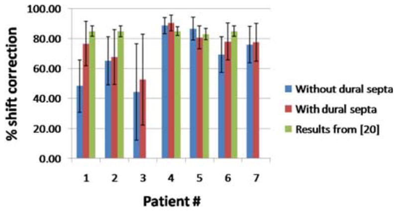
Biomechanical models that describe soft tissue deformation provide a relatively inexpensive way to correct registration errors in image-guided neurosurgical systems caused by nonrigid brain shift. Quantifying the factors that cause this deformation to sufficient precision is a challenging task. To circumvent this difficulty, atlas-based methods have been developed recently that allow for uncertainty, yet still capture the first-order effects associated with deformation. The inverse solution is driven by sparse intraoperative surface measurements, which could bias the reconstruction and affect the subsurface accuracy of the model prediction. Studies using intraoperative MR have shown that the deformation in the midline, tentorium, and contralateral hemisphere is relatively small. The dural septa act as rigid membranes supporting the brain parenchyma and compartmentalizing the brain. Accounting for these structures in models may be an important key to improving subsurface shift accuracy. A novel method to segment the tentorium cerebelli will be described, along with the procedure for modeling the dural septa. Results in seven clinical cases show a qualitative improvement in subsurface shift accuracy making the predicted deformation more congruous with previous observations in the literature. The results also suggest a considerably more important role for hyperosmotic drug modeling for the intraoperative shift correction environment.

摘要

生物力学模型描述了软组织变形,为纠正图像引导神经外科系统中因脑移位导致的非刚性配准误差提供了一种相对廉价的方法。准确量化导致这种变形的因素是一项具有挑战性的任务。为了规避这一困难,最近开发了基于图谱的方法,这些方法允许存在不确定性,但仍能捕捉到与变形相关的一阶效应。逆解由术中稀疏的表面测量驱动,这可能会导致重建偏差,并影响模型预测的亚表面精度。使用术中磁共振成像的研究表明,中线、小脑幕和对侧半球的变形相对较小。硬脑膜隔作为刚性膜支撑脑实质并分隔脑。在模型中考虑这些结构可能是提高亚表面移位精度的重要关键。本文将描述一种新的小脑幕分割方法,以及硬脑膜隔建模的过程。在七个临床病例中的结果表明,亚表面移位精度的定性改善使预测的变形与文献中的先前观察结果更加一致。结果还表明,在术中移位校正环境中,高渗药物建模的作用更为重要。

https://cdn.ncbi.nlm.nih.gov/pmc/blobs/43e0/3864010/4ed10ac24dd6/nihms523852f10.jpg
https://cdn.ncbi.nlm.nih.gov/pmc/blobs/43e0/3864010/89f34f69d062/nihms523852f1.jpg
https://cdn.ncbi.nlm.nih.gov/pmc/blobs/43e0/3864010/e7d6d1ab1a41/nihms523852f2.jpg
https://cdn.ncbi.nlm.nih.gov/pmc/blobs/43e0/3864010/44447abd6aea/nihms523852f3.jpg
https://cdn.ncbi.nlm.nih.gov/pmc/blobs/43e0/3864010/1c6953f91edd/nihms523852f4.jpg
https://cdn.ncbi.nlm.nih.gov/pmc/blobs/43e0/3864010/914eb153e231/nihms523852f5.jpg
https://cdn.ncbi.nlm.nih.gov/pmc/blobs/43e0/3864010/280a439694af/nihms523852f6.jpg
https://cdn.ncbi.nlm.nih.gov/pmc/blobs/43e0/3864010/9b438e48affd/nihms523852f7.jpg
https://cdn.ncbi.nlm.nih.gov/pmc/blobs/43e0/3864010/4b1897d3e918/nihms523852f8.jpg
https://cdn.ncbi.nlm.nih.gov/pmc/blobs/43e0/3864010/6797277324ab/nihms523852f9.jpg
https://cdn.ncbi.nlm.nih.gov/pmc/blobs/43e0/3864010/4ed10ac24dd6/nihms523852f10.jpg

相似文献

1
Intraoperative brain shift compensation: accounting for dural septa.术中脑移位补偿:考虑硬脑膜隔。
IEEE Trans Biomed Eng. 2011 Mar;58(3):499-508. doi: 10.1109/TBME.2010.2093896. Epub 2010 Nov 22.
2
Clinical evaluation of a model-updated image-guidance approach to brain shift compensation: experience in 16 cases.一种模型更新的图像引导脑移位补偿方法的临床评估:16例经验
Int J Comput Assist Radiol Surg. 2016 Aug;11(8):1467-74. doi: 10.1007/s11548-015-1295-x. Epub 2015 Oct 17.
3
Accounting for Deformation in Deep Brain Stimulation Surgery With Models: Comparison to Interventional Magnetic Resonance Imaging.利用模型对深部脑刺激手术中的变形进行定量分析:与介入磁共振成像的比较。
IEEE Trans Biomed Eng. 2020 Oct;67(10):2934-2944. doi: 10.1109/TBME.2020.2974102. Epub 2020 Feb 14.
4
Investigation of intraoperative brain deformation using a 1.5-T interventional MR system: preliminary results.使用1.5-T介入式磁共振系统对术中脑形变的研究:初步结果
IEEE Trans Med Imaging. 1998 Oct;17(5):817-25. doi: 10.1109/42.736050.
5
Intraoperative image updating for brain shift following dural opening.硬脑膜打开后脑移位的术中图像更新。
J Neurosurg. 2017 Jun;126(6):1924-1933. doi: 10.3171/2016.6.JNS152953. Epub 2016 Sep 9.
6
A comparison of thin-plate spline deformation and finite element modeling to compensate for brain shift during tumor resection.薄壳样条变形与有限元建模在肿瘤切除术中补偿脑移位的比较。
Int J Comput Assist Radiol Surg. 2020 Jan;15(1):75-85. doi: 10.1007/s11548-019-02057-2. Epub 2019 Aug 23.
7
Quantification of, visualization of, and compensation for brain shift using intraoperative magnetic resonance imaging.使用术中磁共振成像对脑移位进行量化、可视化及补偿。
Neurosurgery. 2000 Nov;47(5):1070-9; discussion 1079-80. doi: 10.1097/00006123-200011000-00008.
8
Integrating Retraction Modeling Into an Atlas-Based Framework for Brain Shift Prediction.将回缩建模集成到基于图谱的脑移位预测框架中。
IEEE Trans Biomed Eng. 2013 Dec;60(12):3494-504. doi: 10.1109/TBME.2013.2272658. Epub 2013 Jul 10.
9
Evaluation of conoscopic holography for estimating tumor resection cavities in model-based image-guided neurosurgery.基于模型的图像引导神经外科手术中用于估计肿瘤切除腔的锥镜全息术评估
IEEE Trans Biomed Eng. 2014 Jun;61(6):1833-43. doi: 10.1109/TBME.2014.2308299.
10
Estimation of brain deformation for volumetric image updating in protoporphyrin IX fluorescence-guided resection.原卟啉IX荧光引导切除术中用于容积图像更新的脑变形估计
Stereotact Funct Neurosurg. 2010;88(1):1-10. doi: 10.1159/000258143. Epub 2009 Nov 12.

引用本文的文献

1
A position- and time-dependent pressure profile to model viscoelastic mechanical behavior of the brain tissue due to tumor growth.用于模拟肿瘤生长引起的脑组织粘弹性力学行为的位置和时间相关的压力分布。
Comput Methods Biomech Biomed Engin. 2023 May;26(6):660-672. doi: 10.1080/10255842.2022.2082245. Epub 2022 May 31.
2
Impact of brain shift on neural pathways in deep brain stimulation: a preliminary analysis via multi-physics finite element models.脑移位对脑深部刺激神经通路的影响:基于多物理有限元模型的初步分析。
J Neural Eng. 2021 Apr 6;18(5). doi: 10.1088/1741-2552/abf066.
3
A hybrid, image-based and biomechanics-based registration approach to markerless intraoperative nodule localization during video-assisted thoracoscopic surgery.

本文引用的文献

1
Model-Updated Image Guidance: A Statistical Approach to Gravity-Induced Brain Shift.模型更新的图像引导:一种解决重力诱导脑移位的统计方法。
Med Image Comput Comput Assist Interv. 2003 Nov;2878:375-382. doi: 10.1007/978-3-540-39899-8_47.
2
Model-Updated Image-Guided Neurosurgery Using the Finite Element Method: Incorporation of the Falx Cerebri.使用有限元法的模型更新图像引导神经外科手术:大脑镰的纳入
Med Image Comput Comput Assist Interv. 1999 Sep;1679:900-910. doi: 10.1007/10704282_98.
3
A fast and efficient method to compensate for brain shift for tumor resection therapies measured between preoperative and postoperative tomograms.
一种基于图像和生物力学的混合配准方法,用于在电视辅助胸腔镜手术中进行无标记术中结节定位。
Med Image Anal. 2021 Apr;69:101983. doi: 10.1016/j.media.2021.101983. Epub 2021 Jan 30.
4
Accounting for intraoperative brain shift ascribable to cavity collapse during intracranial tumor resection.颅内肿瘤切除术中因腔隙塌陷导致的术中脑移位的计算。
J Med Imaging (Bellingham). 2020 May;7(3):031506. doi: 10.1117/1.JMI.7.3.031506. Epub 2020 Jun 22.
5
Accounting for Deformation in Deep Brain Stimulation Surgery With Models: Comparison to Interventional Magnetic Resonance Imaging.利用模型对深部脑刺激手术中的变形进行定量分析:与介入磁共振成像的比较。
IEEE Trans Biomed Eng. 2020 Oct;67(10):2934-2944. doi: 10.1109/TBME.2020.2974102. Epub 2020 Feb 14.
6
modeling of interstitial pressure in a porcine model: approximation of poroelastic properties and effects of enhanced anatomical structure modeling.猪模型中间隙压力的建模:多孔弹性特性的近似以及增强解剖结构建模的影响
J Med Imaging (Bellingham). 2018 Oct;5(4):045002. doi: 10.1117/1.JMI.5.4.045002. Epub 2018 Dec 6.
7
Falx Cerebri Segmentation via Multi-atlas Boundary Fusion.基于多图谱边界融合的大脑镰分割
Med Image Comput Comput Assist Interv. 2017 Sep;10433:92-99. doi: 10.1007/978-3-319-66182-7_11. Epub 2017 Sep 4.
8
Automatic falx cerebri and tentorium cerebelli segmentation from Magnetic Resonance Images.基于磁共振图像的大脑镰和小脑幕自动分割
Proc SPIE Int Soc Opt Eng. 2017 Feb;10137. doi: 10.1117/12.2255640. Epub 2017 Mar 13.
9
Retrospective study comparing model-based deformation correction to intraoperative magnetic resonance imaging for image-guided neurosurgery.一项比较基于模型的变形校正与术中磁共振成像用于图像引导神经外科手术的回顾性研究。
J Med Imaging (Bellingham). 2017 Jul;4(3):035003. doi: 10.1117/1.JMI.4.3.035003. Epub 2017 Sep 13.
10
Intraoperative Imaging Modalities and Compensation for Brain Shift in Tumor Resection Surgery.肿瘤切除手术中的术中成像模式与脑移位补偿
Int J Biomed Imaging. 2017;2017:6028645. doi: 10.1155/2017/6028645. Epub 2017 Jun 5.
一种快速有效的方法,用于补偿术前和术后断层扫描之间测量的肿瘤切除术治疗中的脑移位。
IEEE Trans Biomed Eng. 2010 Jun;57(6):1285-96. doi: 10.1109/TBME.2009.2039643. Epub 2010 Feb 17.
4
A surface registration method for quantification of intraoperative brain deformations in image-guided neurosurgery.一种用于在图像引导神经外科手术中量化术中脑变形的表面配准方法。
IEEE Trans Inf Technol Biomed. 2009 Nov;13(6):976-83. doi: 10.1109/TITB.2009.2025373. Epub 2009 Jun 19.
5
Semiautomatic registration of pre- and postbrain tumor resection laser range data: method and validation.脑肿瘤切除术前和术后激光测距数据的半自动配准:方法与验证
IEEE Trans Biomed Eng. 2009 Mar;56(3):770-80. doi: 10.1109/TBME.2008.2006758. Epub 2008 Oct 10.
6
Laser range scanning for image-guided neurosurgery: investigation of image-to-physical space registrations.用于图像引导神经外科手术的激光测距扫描:图像到物理空间配准的研究。
Med Phys. 2008 Apr;35(4):1593-605. doi: 10.1118/1.2870216.
7
An atlas-based method to compensate for brain shift: preliminary results.一种基于图谱的脑移位补偿方法:初步结果。
Med Image Anal. 2007 Apr;11(2):128-45. doi: 10.1016/j.media.2006.11.002. Epub 2007 Mar 1.
8
Intraoperative cortical surface characterization using laser range scanning: preliminary results.使用激光测距扫描进行术中皮质表面特征分析:初步结果。
Neurosurgery. 2006 Oct;59(4 Suppl 2):ONS368-76; discussion ONS376-7. doi: 10.1227/01.NEU.0000222665.40301.D2.
9
Patient-specific model of brain deformation: application to medical image registration.个性化脑形变模型:在医学图像配准中的应用。
J Biomech. 2007;40(4):919-29. doi: 10.1016/j.jbiomech.2006.02.021. Epub 2006 May 6.
10
Robust nonrigid registration to capture brain shift from intraoperative MRI.用于捕捉术中磁共振成像引起的脑移位的稳健非刚性配准。
IEEE Trans Med Imaging. 2005 Nov;24(11):1417-27. doi: 10.1109/TMI.2005.856734.