Department of Plastic, Reconstructive and Handsurgery, Klinikum rechts der Isar, Technische Universität München, 80333 München, Germany.
Stem Cells Int. 2010 Dec 1;2011:547247. doi: 10.4061/2011/547247.
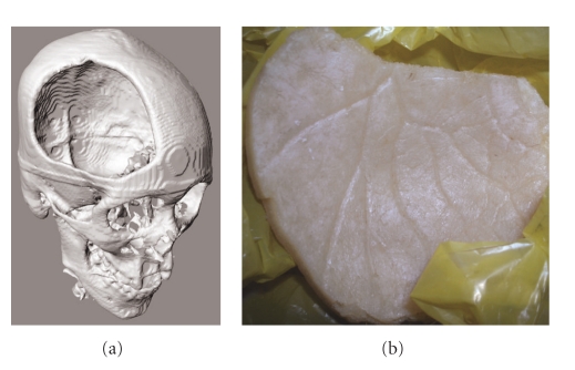
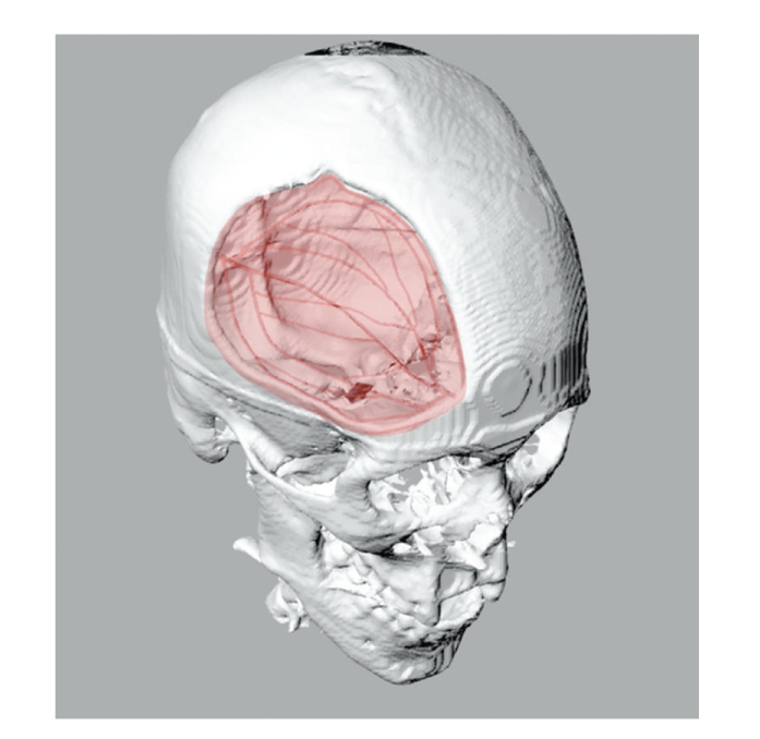
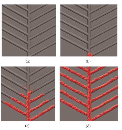
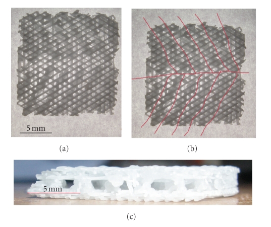
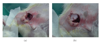
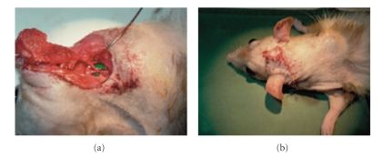
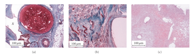
Current tissue engineering techniques are limited by inadequate vascularisation and perfusion of cell-scaffold constructs. Microstructural patterning through biomimetic vascular channels within a polymer scaffold might induce neovascularization, allowing fabrication of large engineered constructs. The network of vascular channels within a frontal-parietal defect in a patient, originating from the anterior branch of the middle meningeal artery, was modeled using computer-aided design (CAD) techniques and subsequently incorporated into polycaprolactone (PCL) scaffolds fabricated using fused deposition modeling (FDM). Bone marrow-derived mesenchymal stem cells (MSCs) were seeded onto the scaffolds and implanted into a rat model, with an arteriovenous bundle inserted at the proximal extent of the vascular network. After 3 weeks, scaffolds were elevated as a prefabricated composite tissue-polymer flap and transferred using microsurgical technique. Histological examination of explanted scaffolds revealed vascular ingrowth along patterned channels, with abundant capillary and connective tissue formation throughout experimental scaffolds, while control scaffolds showed only granulation tissue. All prefabricated constructs transferred as free flaps survived and were viable. We term this concept "vascular guidance," whereby neovascularization is guided through customized channels in a scaffold. Our technique might potentially allow fabrication of much larger tissue-engineered constructs than current technologies allow, as well as allowing tailored construct fabrication with a patient-specific vessel network based on CT scan data and CAD technology.
目前的组织工程技术受到细胞-支架构建体血管化和灌注不足的限制。通过聚合物支架内仿生血管通道进行微观结构图案化可能会诱导新血管生成,从而允许制造大型工程构建体。使用计算机辅助设计 (CAD) 技术对来自脑膜中动脉前支的额顶骨缺损内的血管通道网络进行建模,然后将其整合到使用熔融沉积建模 (FDM) 制造的聚己内酯 (PCL) 支架中。骨髓间充质干细胞 (MSCs) 接种到支架上,并植入大鼠模型中,在血管网络的近端插入动静脉束。3 周后,将支架作为预制复合组织-聚合物皮瓣抬起,并使用显微外科技术转移。对植入的支架进行组织学检查显示,血管沿着图案化通道生长,实验支架中充满了丰富的毛细血管和结缔组织,而对照支架仅显示肉芽组织。所有预制的构建体作为游离皮瓣转移均存活且有活力。我们将这个概念称为“血管引导”,即通过支架中的定制通道引导新血管生成。我们的技术可能允许制造比当前技术允许的更大的组织工程构建体,并且可以根据 CT 扫描数据和 CAD 技术基于患者特定的血管网络来定制构建体制造。