• 文献检索
  • 文档翻译
  • 深度研究
  • 学术资讯
  • Suppr Zotero 插件Zotero 插件
  • 邀请有礼
  • 套餐&价格
  • 历史记录
应用&插件
Suppr Zotero 插件Zotero 插件浏览器插件Mac 客户端Windows 客户端微信小程序
定价
高级版会员购买积分包购买API积分包
服务
文献检索文档翻译深度研究API 文档MCP 服务
关于我们
关于 Suppr公司介绍联系我们用户协议隐私条款
关注我们

Suppr 超能文献

核心技术专利:CN118964589B侵权必究
粤ICP备2023148730 号-1Suppr @ 2026

文献检索

告别复杂PubMed语法,用中文像聊天一样搜索,搜遍4000万医学文献。AI智能推荐,让科研检索更轻松。

立即免费搜索

文件翻译

保留排版,准确专业,支持PDF/Word/PPT等文件格式,支持 12+语言互译。

免费翻译文档

深度研究

AI帮你快速写综述,25分钟生成高质量综述,智能提取关键信息,辅助科研写作。

立即免费体验

应用磁共振成像技术对人源脑胶质瘤球体原位模型进行特征描述。

Characterization of a human tumorsphere glioma orthotopic model using magnetic resonance imaging.

机构信息

Division of Medical Physics, Department of Radiology, The Methodist Hospital-Weill Cornell Medical College and Center for Biotechnology and Informatics, The Methodist Hospital Research Institute, TX 77030, USA.

出版信息

J Neurooncol. 2011 Sep;104(2):473-81. doi: 10.1007/s11060-010-0517-x. Epub 2011 Jan 15.

DOI:10.1007/s11060-010-0517-x
PMID:21240539
原文链接:https://pmc.ncbi.nlm.nih.gov/articles/PMC3161186/
Abstract

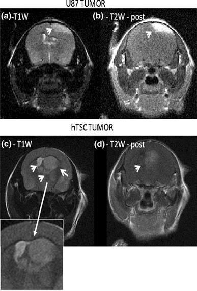
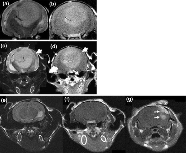
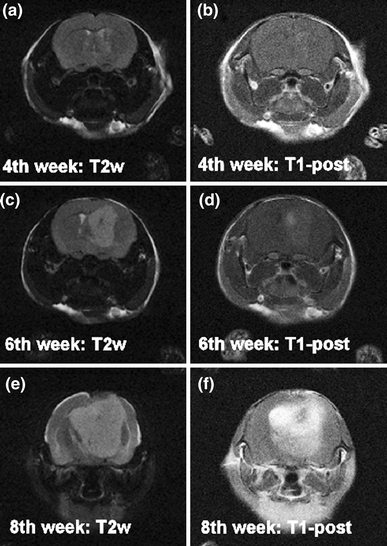
Magnetic resonance imaging (MRI) is the imaging modality of choice by which to monitor patient gliomas and treatment effects, and has been applied to murine models of glioma. However, a major obstacle to the development of effective glioma therapeutics has been that widely used animal models of glioma have not accurately recapitulated the morphological heterogeneity and invasive nature of this very lethal human cancer. This deficiency is being alleviated somewhat as more representative models are being developed, but there is still a clear need for relevant yet practical models that are well-characterized in terms of their MRI features. Hence we sought to chronicle the MRI profile of a recently developed, comparatively straightforward human tumor stem cell (hTSC) derived glioma model in mice using conventional MRI methods. This model reproduces the salient features of gliomas in humans, including florid neoangiogenesis and aggressive invasion of normal brain. Accordingly, the variable, invasive morphology of hTSC gliomas visualized on MRI duplicated that seen in patients, and it differed considerably from the widely used U87 glioma model that does not invade normal brain. After several weeks of tumor growth the hTSC model exhibited an MRI contrast enhancing phenotype having variable intensity and an irregular shape, which mimicked the heterogeneous appearance observed with human glioma patients. The MRI findings reported here support the use of the hTSC glioma xenograft model combined with MRI, as a test platform for assessing candidate therapeutics for glioma, and for developing novel MR methods.

摘要

磁共振成像(MRI)是监测患者脑胶质瘤和治疗效果的首选成像方式,已应用于脑胶质瘤的鼠模型。然而,开发有效的脑胶质瘤治疗方法的一个主要障碍是,广泛使用的脑胶质瘤动物模型未能准确再现这种非常致命的人类癌症的形态异质性和侵袭性。随着更具代表性的模型的开发,这种缺陷在一定程度上得到了缓解,但仍明显需要相关且实用的模型,这些模型在 MRI 特征方面具有良好的特征。因此,我们试图使用常规 MRI 方法来记录最近开发的、相对简单的人类肿瘤干细胞(hTSC)衍生的脑胶质瘤模型在小鼠中的 MRI 特征。该模型再现了人类脑胶质瘤的显著特征,包括丰富的新生血管形成和对正常大脑的侵袭性侵犯。因此,MRI 上可见的 hTSC 脑胶质瘤的可变、侵袭性形态与患者所见的形态相吻合,与广泛使用的不侵犯正常大脑的 U87 脑胶质瘤模型有很大不同。在肿瘤生长数周后,hTSC 模型表现出 MRI 对比增强表型,其强度和形状具有变异性,这与在人类脑胶质瘤患者中观察到的异质性表现相吻合。这里报道的 MRI 结果支持使用 hTSC 脑胶质瘤异种移植模型结合 MRI,作为评估脑胶质瘤候选治疗方法的测试平台,并开发新的 MRI 方法。

https://cdn.ncbi.nlm.nih.gov/pmc/blobs/4bf7/3161186/3ee48d9ea2e5/11060_2010_517_Fig5_HTML.jpg
https://cdn.ncbi.nlm.nih.gov/pmc/blobs/4bf7/3161186/d4272a15ece1/11060_2010_517_Fig1_HTML.jpg
https://cdn.ncbi.nlm.nih.gov/pmc/blobs/4bf7/3161186/45000408e99a/11060_2010_517_Fig2_HTML.jpg
https://cdn.ncbi.nlm.nih.gov/pmc/blobs/4bf7/3161186/effa3d7c803d/11060_2010_517_Fig3_HTML.jpg
https://cdn.ncbi.nlm.nih.gov/pmc/blobs/4bf7/3161186/b22d5458644e/11060_2010_517_Fig4_HTML.jpg
https://cdn.ncbi.nlm.nih.gov/pmc/blobs/4bf7/3161186/3ee48d9ea2e5/11060_2010_517_Fig5_HTML.jpg

相似文献

1
Characterization of a human tumorsphere glioma orthotopic model using magnetic resonance imaging.应用磁共振成像技术对人源脑胶质瘤球体原位模型进行特征描述。
J Neurooncol. 2011 Sep;104(2):473-81. doi: 10.1007/s11060-010-0517-x. Epub 2011 Jan 15.
2
Glioma morphology and tumor-induced vascular alterations revealed in seven rodent glioma models by in vivo magnetic resonance imaging and angiography.通过体内磁共振成像和血管造影术在七种啮齿动物脑胶质瘤模型中揭示的胶质瘤形态和肿瘤诱导的血管改变。
J Magn Reson Imaging. 2010 Aug;32(2):267-75. doi: 10.1002/jmri.22263.
3
Glioma stem cells involved in tumor tissue remodeling in a xenograft model.神经胶质瘤干细胞参与异种移植模型中的肿瘤组织重塑。
J Neurosurg. 2010 Aug;113(2):249-60. doi: 10.3171/2010.2.JNS09335.
4
Iron labeling and pre-clinical MRI visualization of therapeutic human neural stem cells in a murine glioma model.铁标记和临床前 MRI 可视化治疗性人神经干细胞在小鼠胶质瘤模型中的应用。
PLoS One. 2009 Sep 29;4(9):e7218. doi: 10.1371/journal.pone.0007218.
5
[Transplantation and magnetic resonance imaging of transplantable human glioma tissue in the brain of nude mice].[人可移植性脑胶质瘤组织在裸鼠脑内的移植及磁共振成像]
Ai Zheng. 2007 Sep;26(9):937-41.
6
Evaluation of neovascularization patterns in an orthotopic rat glioma model with dynamic contrast-enhanced MRI.利用动态对比增强磁共振成像评估原位大鼠胶质瘤模型中的新生血管形成模式
Acta Radiol. 2017 Sep;58(9):1138-1146. doi: 10.1177/0284185116681038. Epub 2016 Dec 12.
7
The antitumour growth and antiangiogenesis effects of xanthatin in murine glioma dynamically evaluated by dynamic contrast-enhanced magnetic resonance imaging.动态对比增强磁共振成像动态评价莪术呋喃二酮对小鼠脑胶质瘤的抗肿瘤生长和抗血管生成作用。
Phytother Res. 2019 Jan;33(1):149-158. doi: 10.1002/ptr.6207. Epub 2018 Oct 22.
8
Human and rat glioma growth, invasion, and vascularization in a novel chick embryo brain tumor model.新型鸡胚脑肿瘤模型中人类和大鼠胶质瘤的生长、侵袭及血管生成
Clin Exp Metastasis. 2005;22(3):225-36. doi: 10.1007/s10585-005-7889-x.
9
Correlated magnetic resonance imaging and ultramicroscopy (MR-UM) is a tool kit to assess the dynamics of glioma angiogenesis.相关磁共振成像与超微显微镜检查(MR-UM)是一种评估胶质瘤血管生成动态的工具套件。
Elife. 2016 Feb 2;5:e11712. doi: 10.7554/eLife.11712.
10
Development of therapeutics for high grade gliomas using orthotopic rodent models.使用原位啮齿动物模型开发高级别神经胶质瘤的治疗方法。
Curr Med Chem. 2013;20(26):3272-99. doi: 10.2174/0929867311320260011.

引用本文的文献

1
A review of clinical use of surface-enhanced Raman scattering-based biosensing for glioma.基于表面增强拉曼散射的生物传感技术在神经胶质瘤临床应用中的综述。
Front Neurol. 2024 Apr 8;15:1287213. doi: 10.3389/fneur.2024.1287213. eCollection 2024.
2
Characterization of orthotopic xenograft tumor of glioma stem cells (GSCs) on MRI, PET and immunohistochemical staining.胶质瘤干细胞(GSCs)原位异种移植瘤在MRI、PET及免疫组化染色方面的特征
Front Oncol. 2022 Dec 15;12:1085015. doi: 10.3389/fonc.2022.1085015. eCollection 2022.
3
Study on the Function and Mechanism of miR-585-3p Inhibiting the Progression of Ovarian Cancer Cells by Targeting FSCN1 to Block the MAPK Signaling Pathway.

本文引用的文献

1
Activation of tumor cell integrin alphavbeta3 controls angiogenesis and metastatic growth in the brain.肿瘤细胞整合素αvβ3的激活控制着大脑中的血管生成和转移生长。
Proc Natl Acad Sci U S A. 2009 Jun 30;106(26):10666-71. doi: 10.1073/pnas.0903035106. Epub 2009 Jun 16.
2
Imaging glioma cell invasion in vivo reveals mechanisms of dissemination and peritumoral angiogenesis.体内成像神经胶质瘤细胞侵袭揭示了其播散机制和瘤周血管生成。
Glia. 2009 Sep;57(12):1306-15. doi: 10.1002/glia.20850.
3
The role of regulatory T cells in malignant glioma.
miR-585-3p 通过靶向 FSCN1 阻断 MAPK 信号通路抑制卵巢癌细胞进展的功能及机制研究。
Anal Cell Pathol (Amst). 2022 May 13;2022:1732365. doi: 10.1155/2022/1732365. eCollection 2022.
4
Molecular imaging of a fluorescent antibody against epidermal growth factor receptor detects high-grade glioma.荧光抗体靶向表皮生长因子受体的分子成像检测高级别胶质瘤。
Sci Rep. 2021 Mar 11;11(1):5710. doi: 10.1038/s41598-021-84831-4.
5
Clinical implication of cellular vaccine in glioma: current advances and future prospects.细胞疫苗在神经胶质瘤中的临床意义:当前进展与未来展望。
J Exp Clin Cancer Res. 2020 Nov 23;39(1):257. doi: 10.1186/s13046-020-01778-6.
6
MicroRNA-585 inhibits human glioma cell proliferation by directly targeting MDM2.微小RNA-585通过直接靶向MDM2抑制人胶质瘤细胞增殖。
Cancer Cell Int. 2020 Sep 29;20:469. doi: 10.1186/s12935-020-01528-w. eCollection 2020.
7
Patient‑derived orthotopic xenograft glioma models fail to replicate the magnetic resonance imaging features of the original patient tumor.患者来源的原位异种移植脑胶质瘤模型无法复制原始患者肿瘤的磁共振成像特征。
Oncol Rep. 2020 May;43(5):1619-1629. doi: 10.3892/or.2020.7538. Epub 2020 Mar 9.
8
Demonstration of Non-Gaussian Restricted Diffusion in Tumor Cells Using Diffusion Time-Dependent Diffusion-Weighted Magnetic Resonance Imaging Contrast.使用扩散时间依赖性扩散加权磁共振成像对比剂在肿瘤细胞中证明非高斯受限扩散
Front Oncol. 2016 Aug 2;6:179. doi: 10.3389/fonc.2016.00179. eCollection 2016.
9
Establishment and Characterization of a Tumor Stem Cell-Based Glioblastoma Invasion Model.基于肿瘤干细胞的胶质母细胞瘤侵袭模型的建立与表征
PLoS One. 2016 Jul 25;11(7):e0159746. doi: 10.1371/journal.pone.0159746. eCollection 2016.
10
Effect of the JAK2/STAT3 inhibitor SAR317461 on human glioblastoma tumorspheres.JAK2/STAT3抑制剂SAR317461对人胶质母细胞瘤肿瘤球的影响。
J Transl Med. 2015 Aug 18;13:269. doi: 10.1186/s12967-015-0627-5.
调节性T细胞在恶性胶质瘤中的作用。
Anticancer Res. 2008 Mar-Apr;28(2B):1143-50.
4
Glioblastoma multiforme: an emerging paradigm of anti-VEGF therapy.多形性胶质母细胞瘤:抗血管内皮生长因子治疗的新范式
Expert Opin Biol Ther. 2008 Apr;8(4):541-53. doi: 10.1517/14712598.8.4.541.
5
Correlation of F-18-fluoro-ethyl-tyrosin uptake with vascular and cell density in non-contrast-enhancing gliomas.非增强型胶质瘤中F-18-氟乙基酪氨酸摄取与血管及细胞密度的相关性
J Neurooncol. 2008 Jun;88(2):205-10. doi: 10.1007/s11060-008-9551-3.
6
Bevacizumab for recurrent malignant gliomas: efficacy, toxicity, and patterns of recurrence.贝伐单抗治疗复发性恶性胶质瘤:疗效、毒性及复发模式
Neurology. 2008 Mar 4;70(10):779-87. doi: 10.1212/01.wnl.0000304121.57857.38.
7
Paradoxical imaging findings in cerebral gliomas.脑胶质瘤中的矛盾影像学表现。
J Neurol Sci. 2008 Jun 15;269(1-2):180-3. doi: 10.1016/j.jns.2007.12.029. Epub 2008 Feb 5.
8
Genomic changes and gene expression profiles reveal that established glioma cell lines are poorly representative of primary human gliomas.基因组变化和基因表达谱显示,已建立的胶质瘤细胞系不能很好地代表原发性人类胶质瘤。
Mol Cancer Res. 2008 Jan;6(1):21-30. doi: 10.1158/1541-7786.MCR-07-0280. Epub 2008 Jan 9.
9
Chemoradiotherapy in malignant glioma: standard of care and future directions.恶性胶质瘤的放化疗:治疗标准与未来方向
J Clin Oncol. 2007 Sep 10;25(26):4127-36. doi: 10.1200/JCO.2007.11.8554.
10
Standardization of an orthotopic mouse brain tumor model following transplantation of CT-2A astrocytoma cells.CT-2A星形细胞瘤细胞移植后原位小鼠脑肿瘤模型的标准化
Histol Histopathol. 2007 Dec;22(12):1309-26. doi: 10.14670/HH-22.1309.