Laser Microbeam and Medical Program, Beckman Laser Institute and Medical Clinic, 1002 Health Sciences Road, Irvine, CA 92612, USA.
Ann Biomed Eng. 2011 Apr;39(4):1349-57. doi: 10.1007/s10439-011-0269-6. Epub 2011 Feb 19.
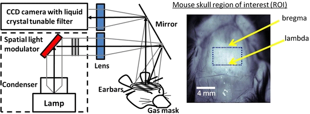
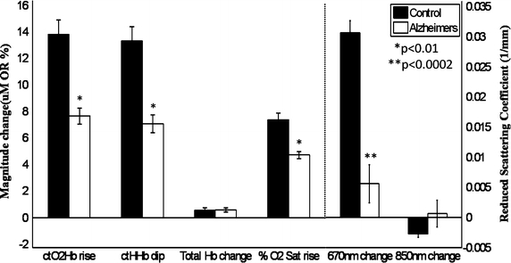
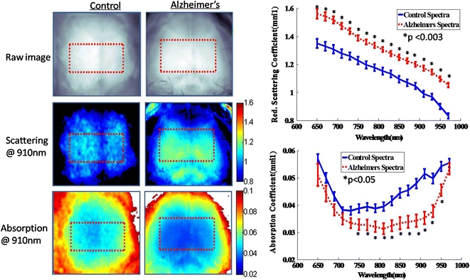
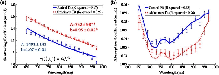
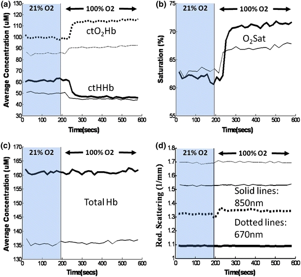
Extensive changes in neural tissue structure and function accompanying Alzheimer's disease (AD) suggest that intrinsic signal optical imaging can provide new contrast mechanisms and insight for assessing AD appearance and progression. In this work, we report the development of a wide-field spatial frequency domain imaging (SFDI) method for non-contact, quantitative in vivo optical imaging of brain tissue composition and function in a triple transgenic mouse AD model (3xTg). SFDI was used to generate optical absorption and scattering maps at up to 17 wavelengths from 650 to 970 nm in 20-month-old 3xTg mice (n = 4) and age-matched controls (n = 6). Wavelength-dependent optical properties were used to form images of tissue hemoglobin (oxy-, deoxy-, and total), oxygen saturation, and water. Significant baseline contrast was observed with 13-26% higher average scattering values and elevated water content (52 ± 2% vs. 31 ± 1%); reduced total tissue hemoglobin content (127 ± 9 μM vs. 174 ± 6 μM); and lower tissue oxygen saturation (57 ± 2% vs. 69 ± 3%) in AD vs. control mice. Oxygen inhalation challenges (100% oxygen) resulted in increased levels of tissue oxy-hemoglobin (ctO(2)Hb) and commensurate reductions in deoxy-hemoglobin (ctHHb), with ~60-70% slower response times and ~7 μM vs. ~14 μM overall changes for 3xTg vs. controls, respectively. Our results show that SFDI is capable of revealing quantitative functional contrast in an AD model and may be a useful method for studying dynamic alterations in AD neural tissue composition and physiology.
伴随阿尔茨海默病(AD)的神经组织结构和功能的广泛变化表明,固有信号光学成像是一种新的对比机制,可以为评估 AD 的出现和进展提供新的见解。在这项工作中,我们报告了一种宽场空间频域成像(SFDI)方法的发展,用于在三转基因 AD 模型(3xTg)中非接触式、定量地对活体脑组织成分和功能进行光学成像。SFDI 用于在 20 个月大的 3xTg 小鼠(n=4)和年龄匹配的对照组(n=6)中从 650nm 到 970nm 生成多达 17 个波长的光吸收和散射图。波长依赖性光学特性用于形成组织血红蛋白(氧合、脱氧和总)、氧饱和度和水的图像。在 AD 小鼠中观察到显著的基线对比,平均散射值高出 13-26%,水含量升高(52±2%比 31±1%);总组织血红蛋白含量降低(127±9μM 比 174±6μM);组织氧饱和度降低(57±2%比 69±3%)。在 AD 小鼠中,氧吸入挑战(100%氧气)导致组织氧合血红蛋白(ctO2Hb)水平升高,同时脱氧血红蛋白(ctHHb)相应降低,3xTg 与对照组相比,反应时间分别慢约 60-70%,总体变化分别约为 7μM 比 14μM。我们的结果表明,SFDI 能够揭示 AD 模型中的定量功能对比,并且可能是研究 AD 神经组织成分和生理学动态变化的有用方法。