Brito Pedro, Penas Susana, Carneiro Angela, Palmares Jorge, Reis F Falcão
Department of Ophthalmology, Hospital S. João, University of Porto, Porto, Portugal.
Case Rep Ophthalmol. 2011 Jan 25;2(1):39-44. doi: 10.1159/000324086.
Syphilis is an infectious disease that can cause a wide variety of ocular signs. One of the rarest manifestations of ocular syphilis is acute syphilitic posterior placoid chorioretinitis (ASPPC). We report on the spectral-domain optical coherence tomography (SD-OCT) features of a case diagnosed with unilateral ASPPC.
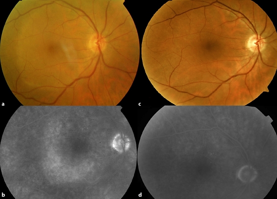
A 64-year-old man presented with a sudden loss of visual acuity (VA) in the right eye. His only clinical sign was a large, geographic, yellow-white lesion centered on the right fovea. Our patient was studied with SD-OCT on presentation and during follow-up, as well as with fluorescein and indocyanine green angiography, electrophysiological study, and serologic and autoimmune screening.
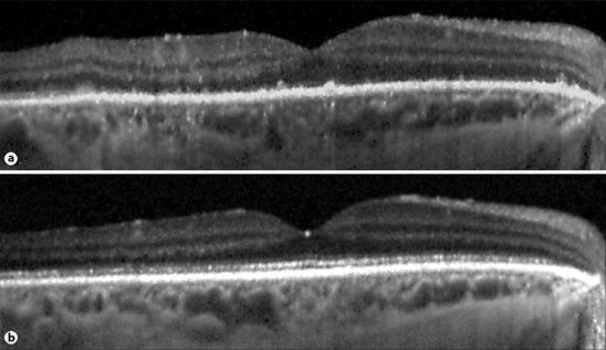
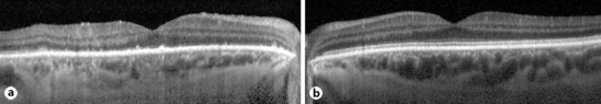
Laboratory workup revealed positive serology for active syphilis and elevated anti-beta2 glycoprotein I antibodies. SD-OCT showed a marked distortion of both the choroidal and outer retinal architecture. After treatment, best-corrected VA improved to 20/25. Pattern electroretinography displayed a severe reduction of P50 amplitude, which improved in late follow-up. Six months after presentation, VA was 20/25 and anti-beta2 glycoprotein I antibodies returned to normal levels.
Our findings are compatible with immunologically mediated temporary physiological impairment of the neuroretina, since the changes seen by SD-OCT could not have normalized if they were due to anatomical injury. The results of our study provide clues to understanding the pathogenesis of this disease and allow us to define a characteristic temporal sequence of events in ASPPC.
梅毒是一种可导致多种眼部体征的传染病。眼部梅毒最罕见的表现之一是急性梅毒性后极部扁平状脉络膜视网膜病变(ASPPC)。我们报告一例单侧ASPPC病例的频域光学相干断层扫描(SD - OCT)特征。
一名64岁男性右眼突然视力下降。其唯一的临床体征是一个以右眼黄斑为中心的大的、地图状、黄白色病变。对该患者在就诊时和随访期间进行了SD - OCT检查,以及荧光素和吲哚菁绿血管造影、电生理检查、血清学和自身免疫筛查。
实验室检查显示活动性梅毒血清学阳性且抗β2糖蛋白I抗体升高。SD - OCT显示脉络膜和视网膜外层结构均有明显扭曲。治疗后,最佳矫正视力提高到20/25。图形视网膜电图显示P50波幅严重降低,在随访后期有所改善。就诊6个月后,视力为20/25,抗β2糖蛋白I抗体恢复到正常水平。
我们的发现与神经视网膜免疫介导的暂时性生理损害相符,因为如果SD - OCT所见的改变是由于解剖损伤,则不可能恢复正常。我们的研究结果为理解该疾病的发病机制提供了线索,并使我们能够确定ASPPC中特征性的时间事件序列。