Department of Radiology, Maastricht University Medical Center, Maastricht, The Netherlands.
PLoS One. 2011 Feb 15;6(2):e17070. doi: 10.1371/journal.pone.0017070.
To prospectively assess the relation between carotid plaque characteristics and the development of new cerebral white matter lesions (WMLs) at MRI.
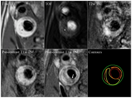
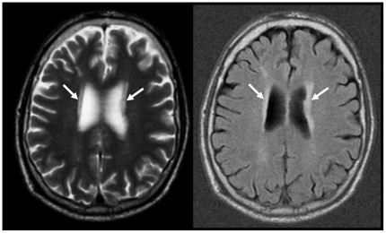
Fifty TIA/stroke patients with ipsilateral 30-69% carotid stenosis underwent MRI of the plaque at baseline. Total plaque volume and markers of vulnerability to thromboembolism (lipid-rich necrotic core [LRNC] volume, fibrous cap [FC] status, and presence of intraplaque hemorrhage [IPH]) were assessed. All patients also underwent brain MRI at baseline and after one year. Ipsilateral cerebral WMLs were quantified with a semiautomatic method.
Mean WML volume significantly increased over a one-year period (6.52 vs. 6.97 mm(3), P = 0.005). WML volume at baseline and WML progression did not significantly differ (P>0.05) between patients with 30-49% and patients with 50-69% stenosis. There was a significant correlation between total plaque volume and baseline ipsilateral WML volume (Spearman ρ = 0.393, P = 0.005). There was no significant correlation between total plaque volume and ipsilateral WML progression. There were no significant associations between LRNC volume and WML volume at baseline and WML progression. WML volume at baseline and WML progression did not significantly differ between patients with a thick and intact FC and patients with a thin and/or ruptured FC. WML volume at baseline and WML progression also did not significantly differ between patients with and without IPH.
The results of this study indicate that carotid plaque burden is significantly associated with WML severity, but that there is no causal relationship between carotid plaque vulnerability and the occurrence of WMLs.
前瞻性评估颈动脉斑块特征与 MRI 上新发脑白质病变(WML)之间的关系。
50 例同侧颈动脉狭窄 30-69%的 TIA/中风患者在基线时接受了斑块 MRI 检查。评估了总斑块体积和易发生血栓栓塞的标志物(富含脂质的坏死核心[LRNC]体积、纤维帽[FC]状态以及斑块内出血[IPH]的存在)。所有患者还在基线和一年后进行了脑部 MRI 检查。采用半自动方法对同侧脑 WML 进行量化。
平均 WML 体积在一年期间显著增加(6.52 与 6.97 mm3,P=0.005)。在狭窄程度为 30-49%和 50-69%的患者之间,基线 WML 体积和 WML 进展之间无显著差异(P>0.05)。总斑块体积与基线同侧 WML 体积之间存在显著相关性(Spearman ρ=0.393,P=0.005)。总斑块体积与同侧 WML 进展之间无显著相关性。LRNC 体积与基线和 WML 进展时的 WML 体积之间无显著关联。在 FC 厚而完整与 FC 薄而/或破裂的患者之间,以及在有和无 IPH 的患者之间,基线 WML 体积和 WML 进展无显著差异。
本研究结果表明,颈动脉斑块负担与 WML 严重程度显著相关,但颈动脉斑块脆弱性与 WML 发生之间无因果关系。