Liu Gangjun, Qi Wenjuan, Yu Lingfeng, Chen Zhongping
Beckman Laser Institute, University of California, Irvine, Irvine, California 92612, USA.
Opt Express. 2011 Feb 14;19(4):3657-66. doi: 10.1364/OE.19.003657.
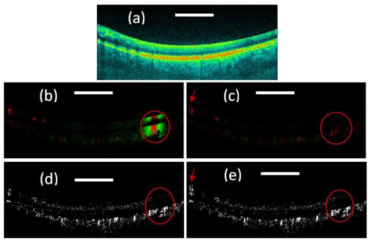
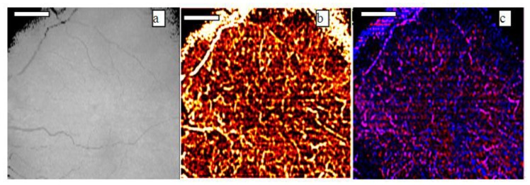
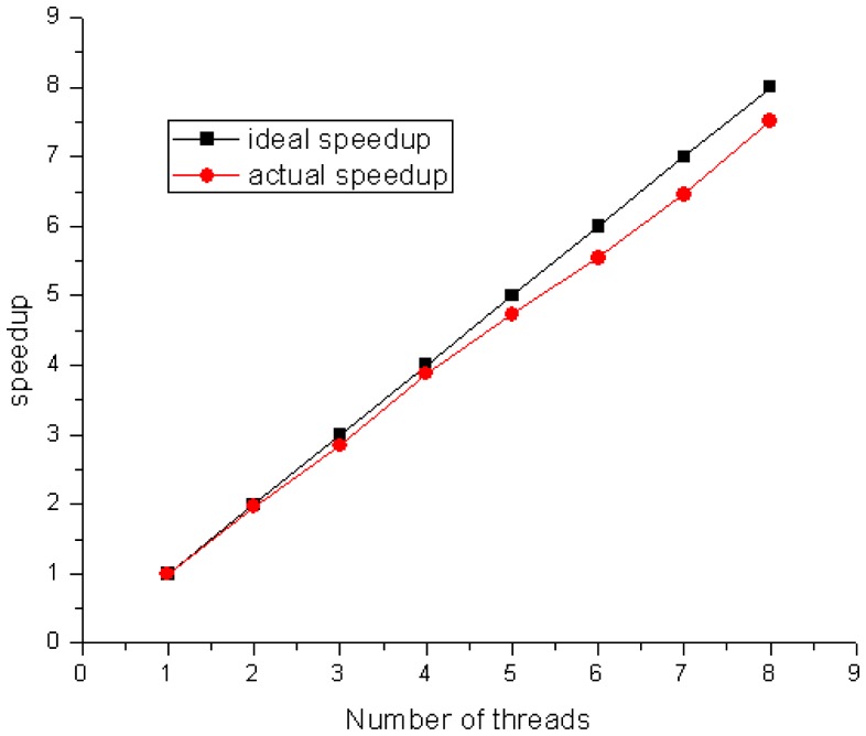
In this paper, we analyze the retinal and choroidal blood vasculature in the posterior segment of the human eye with optimized color Doppler and Doppler variance optical coherence tomography. Depth-resolved structure, color Doppler and Doppler variance images are compared. Blood vessels down to the capillary level were detected and visualized with the optimized optical coherence color Doppler and Doppler variance method. For in-vivo imaging of human eyes, bulk-motion induced bulk phase must be identified and removed before using the color Doppler method. It was found that the Doppler variance method is not sensitive to bulk-motion and the method can be used without correcting the bulk-motion when the sample-movement-induced velocity changes gradually. Real-time processing and displaying of the structure and blood vessel images are very interesting and is demonstrated using a dual quad-core Central Processing Unit (CPU) workstation. High resolution images of choroidal capillary of the vasculature network with phased-resolved color Doppler and Doppler variance are shown.
在本文中,我们使用优化的彩色多普勒和多普勒方差光学相干断层扫描技术,分析人眼后段的视网膜和脉络膜血管系统。比较了深度分辨结构、彩色多普勒和多普勒方差图像。利用优化的光学相干彩色多普勒和多普勒方差方法检测并可视化了直至毛细血管水平的血管。对于人眼的体内成像,在使用彩色多普勒方法之前,必须识别并消除体运动引起的体相位。结果发现,多普勒方差方法对体运动不敏感,当样本运动引起的速度变化逐渐变化时,该方法无需校正体运动即可使用。使用双四核中央处理器(CPU)工作站展示了结构和血管图像的实时处理与显示,这非常有趣。展示了具有相分辨彩色多普勒和多普勒方差的脉管系统网络脉络膜毛细血管的高分辨率图像。