Institute of Clinical Medicine, National Cheng Kung University College of Medicine, Tainan, Taiwan.
J Neuroinflammation. 2011 Apr 25;8:40. doi: 10.1186/1742-2094-8-40.
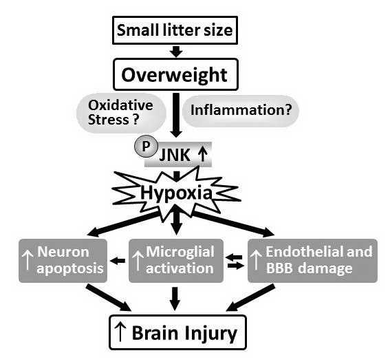
Apoptosis, neuroinflammation and blood-brain barrier (BBB) damage affect the susceptibility of the developing brain to hypoxic-ischemic (HI) insults. c-Jun N-terminal kinase (JNK) is an important mediator of insulin resistance in obesity. We hypothesized that neonatal overweight aggravates HI brain damage through JNK hyperactivation-mediated upregulation of neuronal apoptosis, neuroinflammation and BBB leakage in rat pups.
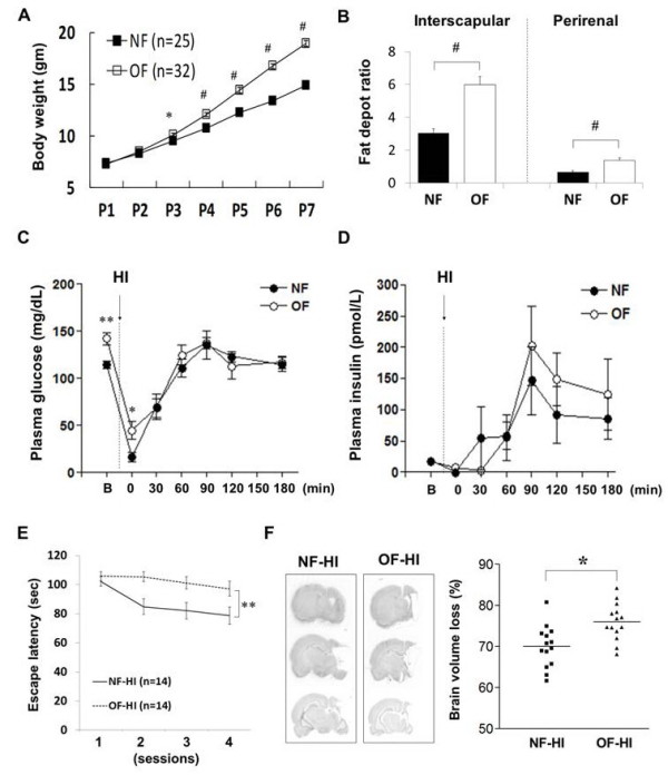
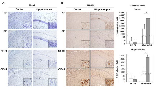
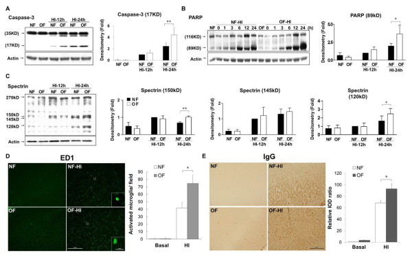
Overweight (OF) pups were established by reducing the litter size to 6, and control (NF) pups by keeping the litter size at 12 from postnatal (P) day 1 before HI on P7. Immunohistochemistry and immunoblotting were used to determine the TUNEL-(+) cells and BBB damage, cleaved caspase-3 and poly (ADP-ribose) polymerase (PARP), and phospho-JNK and phospho-BimEL levels. Immunofluorescence was performed to determine the cellular distribution of phospho-JNK.
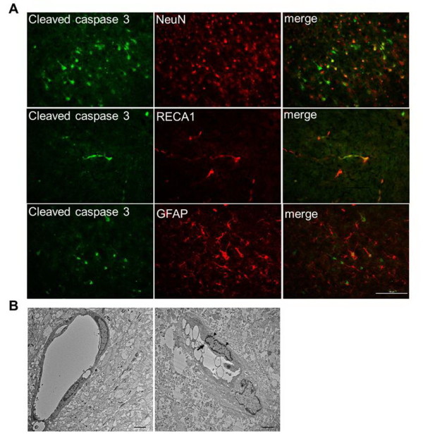
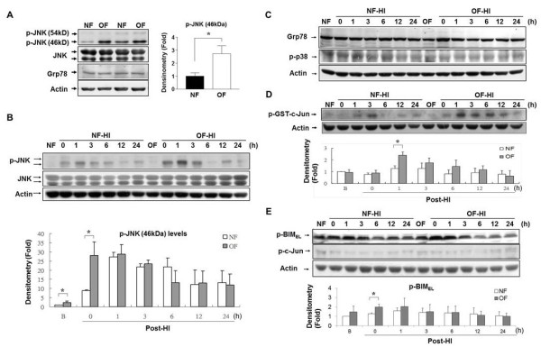
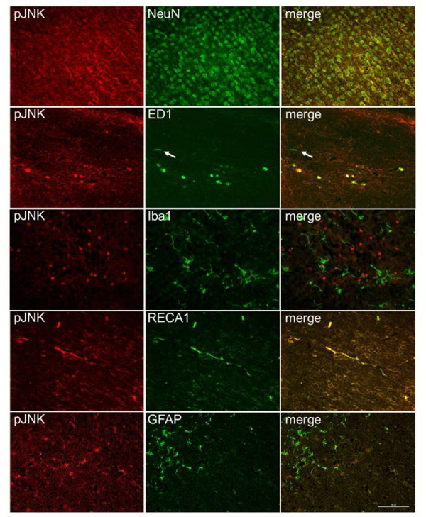
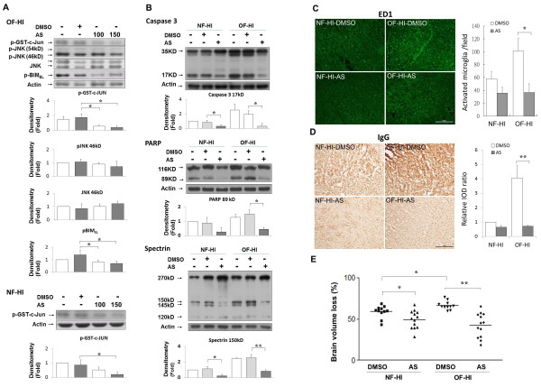
Compared with NF pups, OF pups had a significantly heavier body-weight and greater fat deposition on P7. Compared with the NF-HI group, the OF-HI group showed significant increases of TUNEL-(+) cells, cleaved levels of caspase-3 and PARP, and ED1-(+) activated microglia and BBB damage in the cortex 24 hours post-HI. Immunofluorescence of the OF-HI pups showed that activated-caspase 3 expression was found mainly in NeuN-(+) neurons and RECA1-(+) vascular endothelial cells 24 hours post-HI. The OF-HI group also had prolonged escape latency in the Morris water maze test and greater brain-volume loss compared with the NF-HI group when assessed at adulthood. Phospho-JNK and phospho-BimEL levels were higher in OF-HI pups than in NF-HI pups immediately post-HI. JNK activation in OF-HI pups was mainly expressed in neurons, microglia and vascular endothelial cells. Inhibiting JNK activity by AS601245 caused more attenuation of cleaved caspase-3 and PARP, a greater reduction of microglial activation and BBB damage post-HI, and significantly reduced brain damage in OF-HI than in NF-HI pups.
Neonatal overweight increased HI-induced neuronal apoptosis, microglial activation and BBB damage, and aggravated HI brain damage in rat pups through JNK hyperactivation.
细胞凋亡、神经炎症和血脑屏障(BBB)损伤会影响发育中大脑对缺氧缺血(HI)损伤的易感性。c-Jun N 端激酶(JNK)是肥胖症中胰岛素抵抗的重要介质。我们假设,新生儿超重会通过 JNK 过度激活介导的神经元凋亡、神经炎症和 BBB 渗漏增加,加重 HI 脑损伤。
通过在 HI 前第 7 天减少每个窝仔数至 6 只来建立超重(OF)仔鼠,通过保持每个窝仔数在 12 只来建立对照(NF)仔鼠。免疫组化和免疫印迹用于确定 TUNEL-(+)细胞和 BBB 损伤、cleaved caspase-3 和多聚(ADP-核糖)聚合酶(PARP),以及磷酸化-JNK 和磷酸化-BimEL 水平。免疫荧光用于确定磷酸化-JNK 的细胞分布。
与 NF 仔鼠相比,OF 仔鼠在 P7 时体重明显增加,脂肪沉积更多。与 NF-HI 组相比,OF-HI 组在 HI 后 24 小时,皮层中 TUNEL-(+)细胞、cleaved caspase-3 和 PARP 的水平、ED1-(+)激活的小胶质细胞和 BBB 损伤均显著增加。OF-HI 仔鼠的免疫荧光显示,激活的 caspase-3 表达主要在 NeuN-(+)神经元和 RECA1-(+)血管内皮细胞中,HI 后 24 小时发现。与 NF-HI 组相比,OF-HI 组在成年后在 Morris 水迷宫测试中表现出更长的逃避潜伏期和更大的脑容量损失。OF-HI 仔鼠中磷酸化-JNK 和磷酸化-BimEL 的水平高于 NF-HI 仔鼠。HI 后,OF-HI 仔鼠中的 JNK 激活主要在神经元、小胶质细胞和血管内皮细胞中表达。通过 AS601245 抑制 JNK 活性可使 HI 后 cleaved caspase-3 和 PARP 的减少更为明显,小胶质细胞激活和 BBB 损伤的减少更为明显,与 NF-HI 仔鼠相比,OF-HI 仔鼠的脑损伤明显减少。
新生儿超重通过 JNK 过度激活增加 HI 诱导的神经元凋亡、小胶质细胞激活和 BBB 损伤,加重了 HI 脑损伤。