Department of Nuclear Medicine, The Queen Elizabeth Hospital, Adelaide, South Australia.
NMR Biomed. 2011 Dec;24(10):1302-12. doi: 10.1002/nbm.1692. Epub 2011 May 11.
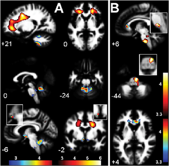
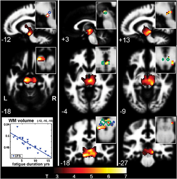
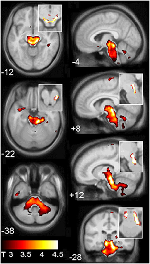
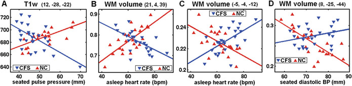
To explore brain involvement in chronic fatigue syndrome (CFS), the statistical parametric mapping of brain MR images has been extended to voxel-based regressions against clinical scores. Using SPM5 we performed voxel-based morphometry (VBM) and analysed T(1) - and T(2) -weighted spin-echo MR signal levels in 25 CFS subjects and 25 normal controls (NC). Clinical scores included CFS fatigue duration, a score based on the 10 most common CFS symptoms, the Bell score, the hospital anxiety and depression scale (HADS) anxiety and depression, and hemodynamic parameters from 24-h blood pressure monitoring. We also performed group × hemodynamic score interaction regressions to detect locations where MR regressions were opposite for CFS and NC, thereby indicating abnormality in the CFS group. In the midbrain, white matter volume was observed to decrease with increasing fatigue duration. For T(1) -weighted MR and white matter volume, group × hemodynamic score interactions were detected in the brainstem [strongest in midbrain grey matter (GM)], deep prefrontal white matter (WM), the caudal basal pons and hypothalamus. A strong correlation in CFS between brainstem GM volume and pulse pressure suggested impaired cerebrovascular autoregulation. It can be argued that at least some of these changes could arise from astrocyte dysfunction. These results are consistent with an insult to the midbrain at fatigue onset that affects multiple feedback control loops to suppress cerebral motor and cognitive activity and disrupt local CNS homeostasis, including resetting of some elements of the autonomic nervous system (ANS).
为了探究慢性疲劳综合征(CFS)患者的大脑受累情况,我们将脑磁共振图像的统计参数图分析方法扩展到了针对临床评分的体素回归分析。我们使用 SPM5 进行了基于体素的形态计量学(VBM)分析,并分析了 25 例 CFS 患者和 25 例正常对照(NC)的 T1 和 T2 加权自旋回波磁共振信号水平。临床评分包括 CFS 疲劳持续时间、基于 10 种最常见 CFS 症状的评分、Bell 评分、医院焦虑和抑郁量表(HADS)焦虑和抑郁评分以及 24 小时血压监测的血液动力学参数。我们还进行了组×血液动力学评分交互回归分析,以检测 CFS 和 NC 组的磁共振回归相反的位置,从而表明 CFS 组存在异常。在中脑,观察到随着疲劳持续时间的增加,白质体积减少。对于 T1 加权磁共振和白质体积,在脑干中检测到组×血液动力学评分的相互作用(在中脑灰质(GM)最强)、深部前额叶白质(WM)、尾部基底脑桥和下丘脑。CFS 患者中脑干 GM 体积与脉压之间的强烈相关性表明脑血管自动调节受损。可以认为,这些变化至少有一部分可能是由于星形胶质细胞功能障碍引起的。这些结果与疲劳发作时中脑受到损伤的观点一致,这会影响多个反馈控制回路,抑制大脑的运动和认知活动,并破坏局部中枢神经系统的内稳态,包括自主神经系统(ANS)的某些元素的重置。