Shan Zack Y, Kwiatek Richard, Burnet Richard, Del Fante Peter, Staines Donald R, Marshall-Gradisnik Sonya M, Barnden Leighton R
National Centre for Neuroimmunology and Emerging Diseases, Menzies Health Institute Queensland, Griffith University, Southport, Australia.
Division of Medical Subspecialities, Lyell McEwin Hospital, Elizabeth Vale, SA, Australia.
J Magn Reson Imaging. 2016 Nov;44(5):1301-1311. doi: 10.1002/jmri.25283. Epub 2016 Apr 28.
To examine progressive brain changes associated with chronic fatigue syndrome (CFS).
We investigated progressive brain changes with longitudinal MRI in 15 CFS and 10 normal controls (NCs) scanned twice 6 years apart on the same 1.5 Tesla (T) scanner. MR images yielded gray matter (GM) volumes, white matter (WM) volumes, and T1- and T2-weighted signal intensities (T1w and T2w). Each participant was characterized with Bell disability scores, and somatic and neurological symptom scores. We tested for differences in longitudinal changes between CFS and NC groups, inter group differences between pooled CFS and pooled NC populations, and correlations between MRI and symptom scores using voxel based morphometry. The analysis methodologies were first optimized using simulated atrophy.
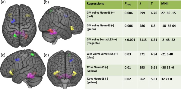
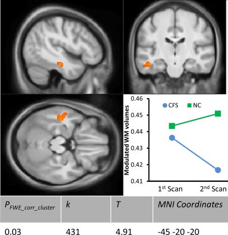
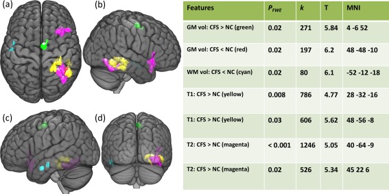
We found a significant decrease in WM volumes in the left inferior fronto-occipital fasciculus (IFOF) in CFS while in NCs it was unchanged (family wise error adjusted cluster level P value, P < 0.05). This longitudinal finding was consolidated by the group comparisons which detected significantly decreased regional WM volumes in adjacent regions (P < 0.05) and decreased GM and blood volumes in contralateral regions (P < 0.05). Moreover, the regional GM and WM volumes and T2w in those areas showed significant correlations with CFS symptom scores (P < 0.05).
The results suggested that CFS is associated with IFOF WM deficits which continue to deteriorate at an abnormal rate. J. Magn. Reson. Imaging 2016;44:1301-1311.
研究与慢性疲劳综合征(CFS)相关的脑部进行性变化。
我们对15名CFS患者和10名正常对照者(NCs)进行了纵向MRI研究,在同一台1.5特斯拉(T)扫描仪上相隔6年进行了两次扫描。MR图像得出灰质(GM)体积、白质(WM)体积以及T1加权和T2加权信号强度(T1w和T2w)。每位参与者都用贝尔残疾评分以及躯体和神经症状评分进行了特征描述。我们使用基于体素的形态测量法测试了CFS组和NC组之间纵向变化的差异、合并的CFS和合并的NC人群之间的组间差异以及MRI与症状评分之间的相关性。首先使用模拟萎缩对分析方法进行了优化。
我们发现CFS患者左侧额枕下束(IFOF)的WM体积显著减少,而NCs则无变化(家族性错误校正聚类水平P值,P<0.05)。这一纵向发现通过组间比较得到了巩固,该比较检测到相邻区域的区域WM体积显著减少(P<0.05),对侧区域的GM和血容量减少(P<0.05)。此外,这些区域的区域GM和WM体积以及T2w与CFS症状评分显示出显著相关性(P<0.05)。
结果表明,CFS与IFOF的WM缺陷有关,该缺陷以异常速率持续恶化。《磁共振成像杂志》2016年;44:1301 - 1311。