• 文献检索
  • 文档翻译
  • 深度研究
  • 学术资讯
  • Suppr Zotero 插件Zotero 插件
  • 邀请有礼
  • 套餐&价格
  • 历史记录
应用&插件
Suppr Zotero 插件Zotero 插件浏览器插件Mac 客户端Windows 客户端微信小程序
定价
高级版会员购买积分包购买API积分包
服务
文献检索文档翻译深度研究API 文档MCP 服务
关于我们
关于 Suppr公司介绍联系我们用户协议隐私条款
关注我们

Suppr 超能文献

核心技术专利:CN118964589B侵权必究
粤ICP备2023148730 号-1Suppr @ 2026

文献检索

告别复杂PubMed语法,用中文像聊天一样搜索,搜遍4000万医学文献。AI智能推荐,让科研检索更轻松。

立即免费搜索

文件翻译

保留排版,准确专业,支持PDF/Word/PPT等文件格式,支持 12+语言互译。

免费翻译文档

深度研究

AI帮你快速写综述,25分钟生成高质量综述,智能提取关键信息,辅助科研写作。

立即免费体验

通过使用透射电子断层扫描技术来确定比表面积,以对纳米材料进行表征和定义。

Determination of the volume-specific surface area by using transmission electron tomography for characterization and definition of nanomaterials.

机构信息

EM-unit, CODA-CERVA, Groeselenberg 99, Brussels, Belgium.

出版信息

J Nanobiotechnology. 2011 May 11;9:17. doi: 10.1186/1477-3155-9-17.

DOI:10.1186/1477-3155-9-17
PMID:21569366
原文链接:https://pmc.ncbi.nlm.nih.gov/articles/PMC3113930/
Abstract

BACKGROUND

Transmission electron microscopy (TEM) remains an important technique to investigate the size, shape and surface characteristics of particles at the nanometer scale. Resulting micrographs are two dimensional projections of objects and their interpretation can be difficult. Recently, electron tomography (ET) is increasingly used to reveal the morphology of nanomaterials (NM) in 3D. In this study, we examined the feasibility to visualize and measure silica and gold NM in suspension using conventional bright field electron tomography.

RESULTS

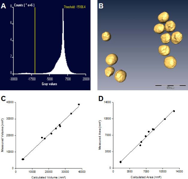
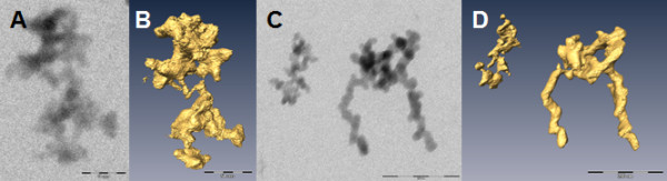
The general morphology of gold and silica NM was visualized in 3D by conventional TEM in bright field mode. In orthoslices of the examined NM the surface features of a NM could be seen and measured without interference of higher or lower lying structures inherent to conventional TEM. Segmentation by isosurface rendering allowed visualizing the 3D information of an electron tomographic reconstruction in greater detail than digital slicing. From the 3D reconstructions, the surface area and the volume of the examined NM could be estimated directly and the volume-specific surface area (VSSA) was calculated. The mean VSSA of all examined NM was significantly larger than the threshold of 60 m(2)/cm(3). The high correlation between the measured values of area and volume gold nanoparticles with a known spherical morphology and the areas and volumes calculated from the equivalent circle diameter (ECD) of projected nanoparticles (NP) indicates that the values measured from electron tomographic reconstructions are valid for these gold particles.

CONCLUSION

The characterization and definition of the examined gold and silica NM can benefit from application of conventional bright field electron tomography: the NM can be visualized in 3D, while surface features and the VSSA can be measured.

摘要

背景

透射电子显微镜(TEM)仍然是一种研究纳米尺度颗粒尺寸、形状和表面特征的重要技术。得到的显微照片是物体的二维投影,其解释可能很困难。最近,电子断层扫描(ET)越来越多地用于揭示纳米材料(NM)的 3D 形态。在这项研究中,我们考察了使用常规明场电子断层扫描可视化和测量悬浮液中二氧化硅和金 NM 的可行性。

结果

通过常规 TEM 明场模式在 3D 中可视化了金和二氧化硅 NM 的一般形态。在检查 NM 的正交切片中,可以看到 NM 的表面特征,并且可以进行测量,而不会受到常规 TEM 中固有更高或更低层结构的干扰。通过等表面渲染进行分割,可以比数字切片更详细地可视化电子断层重建的 3D 信息。从 3D 重建中,可以直接估计检查 NM 的表面积和体积,并计算体积比表面积(VSSA)。所有检查 NM 的平均 VSSA 明显大于 60 m²/cm³的阈值。具有已知球形形态的测量值与投影纳米粒子(NP)的等效圆直径(ECD)计算出的面积和体积之间的高度相关性表明,从电子断层重建中测量的值对这些金粒子有效。

结论

常规明场电子断层扫描的应用可以使所检查的金和二氧化硅 NM 的特征和定义受益:可以在 3D 中可视化 NM,同时可以测量表面特征和 VSSA。

https://cdn.ncbi.nlm.nih.gov/pmc/blobs/68fb/3113930/3c22e5d21c73/1477-3155-9-17-3.jpg
https://cdn.ncbi.nlm.nih.gov/pmc/blobs/68fb/3113930/b356c0237601/1477-3155-9-17-1.jpg
https://cdn.ncbi.nlm.nih.gov/pmc/blobs/68fb/3113930/501667394c8a/1477-3155-9-17-2.jpg
https://cdn.ncbi.nlm.nih.gov/pmc/blobs/68fb/3113930/3c22e5d21c73/1477-3155-9-17-3.jpg
https://cdn.ncbi.nlm.nih.gov/pmc/blobs/68fb/3113930/b356c0237601/1477-3155-9-17-1.jpg
https://cdn.ncbi.nlm.nih.gov/pmc/blobs/68fb/3113930/501667394c8a/1477-3155-9-17-2.jpg
https://cdn.ncbi.nlm.nih.gov/pmc/blobs/68fb/3113930/3c22e5d21c73/1477-3155-9-17-3.jpg

相似文献

1
Determination of the volume-specific surface area by using transmission electron tomography for characterization and definition of nanomaterials.通过使用透射电子断层扫描技术来确定比表面积,以对纳米材料进行表征和定义。
J Nanobiotechnology. 2011 May 11;9:17. doi: 10.1186/1477-3155-9-17.
2
A new approach for 3D reconstruction from bright field TEM imaging: beam precession assisted electron tomography.一种新的明场 TEM 成像三维重构方法:进动辅助电子断层扫描。
Ultramicroscopy. 2011 Aug-Oct;111(9-10):1504-11. doi: 10.1016/j.ultramic.2011.06.002. Epub 2011 Jun 30.
3
Morphology visualization of irregular shape bacteria by electron holography and tomography.通过电子全息术和断层扫描对不规则形状细菌进行形态学可视化
Microsc Res Tech. 2017 Dec;80(12):1249-1255. doi: 10.1002/jemt.22923. Epub 2017 Aug 11.
4
Electron tomography in plant cell biology.植物细胞生物学中的电子断层扫描技术。
Microscopy (Oxf). 2019 Feb 1;68(1):69-79. doi: 10.1093/jmicro/dfy133.
5
Three-dimensional reconstruction of nucleolar components by electron microscope tomography.通过电子显微镜断层扫描对核仁成分进行三维重建。
Methods Mol Biol. 2008;463:137-58. doi: 10.1007/978-1-59745-406-3_10.
6
Reconstructing plant cells in 3D by serial section electron tomography.通过连续切片电子断层扫描技术对植物细胞进行三维重建。
Methods Mol Biol. 2014;1080:159-70. doi: 10.1007/978-1-62703-643-6_13.
7
Formation and thermal stability of gold-silica nanohybrids: insight into the mechanism and morphology by electron tomography.金-硅纳米杂化材料的形成与热稳定性:通过电子断层扫描技术深入了解其形成机制与形态。
Angew Chem Int Ed Engl. 2014 Apr 7;53(15):3970-4. doi: 10.1002/anie.201309288. Epub 2014 Mar 5.
8
Cryo-electron microscopy and cryo-electron tomography of nanoparticles.纳米颗粒的冷冻电子显微镜和冷冻电子断层扫描。
Wiley Interdiscip Rev Nanomed Nanobiotechnol. 2017 Mar;9(2). doi: 10.1002/wnan.1417. Epub 2016 Jun 23.
9
Three-dimensional morphology of iron oxide nanoparticles with reactive concave surfaces. A compressed sensing-electron tomography (CS-ET) approach.具有反应性凹面的氧化铁纳米粒子的三维形态。一种压缩感知-电子断层扫描(CS-ET)方法。
Nano Lett. 2011 Nov 9;11(11):4666-73. doi: 10.1021/nl202253a. Epub 2011 Oct 5.
10
Comparison of 3D cellular imaging techniques based on scanned electron probes: Serial block face SEM vs. Axial bright-field STEM tomography.基于扫描电子探针的 3D 细胞成像技术比较:连续块面扫描电镜与轴向明场扫描透射电镜断层成像。
J Struct Biol. 2018 Jun;202(3):216-228. doi: 10.1016/j.jsb.2018.01.012. Epub 2018 Feb 1.

引用本文的文献

1
Using Femtosecond Laser Light to Investigate the Concentration- and Size-Dependent Nonlinear Optical Properties of Laser-Ablated CuO Quantum Dots.利用飞秒激光研究激光烧蚀法制备的CuO量子点浓度和尺寸依赖的非线性光学性质。
Nanomaterials (Basel). 2024 Oct 18;14(20):1674. doi: 10.3390/nano14201674.
2
Advanced materials and technologies for oral diseases.口腔疾病的先进材料与技术
Sci Technol Adv Mater. 2023 Jan 4;24(1):2156257. doi: 10.1080/14686996.2022.2156257. eCollection 2023.
3
Using Femtosecond Laser Pulses to Explore the Nonlinear Optical Properties of Au NP Colloids That Were Synthesized by Laser Ablation.

本文引用的文献

1
Quantification of metallic nanoparticle morphology on TiO2 using HAADF-STEM tomography.使用 HAADF-STEM 断层扫描技术定量 TiO2 上的金属纳米颗粒形态。
Ultramicroscopy. 2010 Aug;110(9):1120-7. doi: 10.1016/j.ultramic.2010.04.003. Epub 2010 Apr 18.
2
High-performance iterative electron tomography reconstruction with long-object compensation using graphics processing units (GPUs).使用图形处理单元 (GPU) 进行具有长对象补偿的高性能迭代电子断层重建。
J Struct Biol. 2010 Aug;171(2):142-53. doi: 10.1016/j.jsb.2010.03.018. Epub 2010 Apr 4.
3
Three-dimensional analysis of carbon nanotube networks in interconnects by electron tomography without missing wedge artifacts.
利用飞秒激光脉冲探究通过激光烧蚀合成的金纳米粒子胶体的非线性光学性质。
Nanomaterials (Basel). 2022 Aug 28;12(17):2980. doi: 10.3390/nano12172980.
4
Parakeet: a digital twin software pipeline to assess the impact of experimental parameters on tomographic reconstructions for cryo-electron tomography.虎皮鹦鹉:一种用于评估实验参数对冷冻电子断层扫描层析重建影响的数字孪生软件管道。
Open Biol. 2021 Oct;11(10):210160. doi: 10.1098/rsob.210160. Epub 2021 Oct 27.
5
Toxicity of gold nanoparticles (AuNPs): A review.金纳米颗粒(AuNPs)的毒性:综述
Biochem Biophys Rep. 2021 Apr 10;26:100991. doi: 10.1016/j.bbrep.2021.100991. eCollection 2021 Jul.
6
Metal nanomaterials: Immune effects and implications of physicochemical properties on sensitization, elicitation, and exacerbation of allergic disease.金属纳米材料:免疫效应及理化性质对变应性疾病致敏、激发和加重的影响。
J Immunotoxicol. 2019 Dec;16(1):87-124. doi: 10.1080/1547691X.2019.1605553.
7
Physico-chemical properties of manufactured nanomaterials - Characterisation and relevant methods. An outlook based on the OECD Testing Programme.纳米材料的物理化学特性 - 特性描述和相关方法。基于经合组织测试计划的展望。
Regul Toxicol Pharmacol. 2018 Feb;92:8-28. doi: 10.1016/j.yrtph.2017.10.019. Epub 2017 Oct 23.
8
Reliable nanomaterial classification of powders using the volume-specific surface area method.使用比容表面积法对粉末进行可靠的纳米材料分类。
J Nanopart Res. 2017;19(2):61. doi: 10.1007/s11051-017-3741-x. Epub 2017 Feb 11.
9
Volume determination of irregularly-shaped quasi-spherical nanoparticles.不规则准球形纳米粒子的体积测定。
Anal Bioanal Chem. 2016 Nov;408(28):7897-7903. doi: 10.1007/s00216-016-9909-x. Epub 2016 Sep 22.
10
Morphogenesis of pestiviruses: new insights from ultrastructural studies of strain Giraffe-1.瘟病毒的形态发生:来自长颈鹿-1株超微结构研究的新见解
J Virol. 2014 Mar;88(5):2717-24. doi: 10.1128/JVI.03237-13. Epub 2013 Dec 18.
通过电子断层扫描技术对互连线中的碳纳米管网络进行三维分析,避免了缺失楔形伪影。
Microsc Microanal. 2010 Apr;16(2):210-7. doi: 10.1017/S1431927609991371. Epub 2010 Feb 26.
4
Comparing electron tomography and HRTEM slicing methods as tools to measure the thickness of nanoparticles.比较电子断层扫描和高分辨率透射电子显微镜切片方法作为测量纳米颗粒厚度的工具。
Ultramicroscopy. 2009 Jun;109(7):788-96. doi: 10.1016/j.ultramic.2009.02.002. Epub 2009 Feb 28.
5
Electron tomography for heterogeneous catalysts and related nanostructured materials.用于多相催化剂及相关纳米结构材料的电子断层扫描技术
Chem Rev. 2009 May;109(5):1613-29. doi: 10.1021/cr800434t.
6
Optimal threshold selection for tomogram segmentation by projection distance minimization.通过投影距离最小化进行断层图像分割的最优阈值选择
IEEE Trans Med Imaging. 2009 May;28(5):676-86. doi: 10.1109/TMI.2008.2010437. Epub 2008 Dec 9.
7
3D imaging of nanomaterials by discrete tomography.通过离散断层扫描对纳米材料进行3D成像。
Ultramicroscopy. 2009 May;109(6):730-40. doi: 10.1016/j.ultramic.2009.01.009. Epub 2009 Jan 31.
8
Electron tomography of negatively stained complex viruses: application in their diagnosis.负染复杂病毒的电子断层成像:在其诊断中的应用。
Diagn Pathol. 2009 Feb 10;4:5. doi: 10.1186/1746-1596-4-5.
9
Nanoparticle shape and configuration analysis by transmission electron tomography.通过透射电子断层扫描进行纳米颗粒形状和结构分析。
J Microsc. 2008 Jun;230(Pt 3):382-7. doi: 10.1111/j.1365-2818.2008.01996.x.
10
Nanoparticle analysis and characterization methodologies in environmental risk assessment of engineered nanoparticles.工程纳米颗粒环境风险评估中的纳米颗粒分析与表征方法
Ecotoxicology. 2008 Jul;17(5):344-61. doi: 10.1007/s10646-008-0225-x. Epub 2008 May 16.