Cao Y Z, Friedman-Kien A E, Huang Y X, Li X L, Mirabile M, Moudgil T, Zucker-Franklin D, Ho D D
Department of Microbiology, New York University Medical Center, New York 10016.
J Virol. 1990 Jun;64(6):2553-9. doi: 10.1128/JVI.64.6.2553-2559.1990.
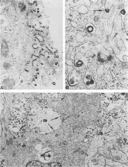
Five hepatoma cell lines, including CZHC/8571, PLC/PRF/5, Hep3B, HepG2, and HUH7, were inoculated with three diverse isolates of human immunodeficiency virus type 1 (HIV-1). Productive infection was noted in all hepatoma cell lines, and expression of viral p24 antigen lasted for over 3 months, but its level decreased in proportion to the number of viable cells. HIV-1 antigens were also found in the cells by immunohistochemical staining and radioimmunoprecipitation assay, as were viral RNA by in situ hybridization and HIV-1-like particles by electron microscopy. Virus yield assays were also positive on supernatant fluids collected from hepatoma cultures inoculated with HIV-1. Despite their susceptibility to infection, all five hepatoma cell lines were negative for CD4 by immunofluorescence and for CD4 mRNA by slot-blot hybridization. In addition, HIV-1 infection of hepatoma cell lines was not blocked by anti-CD4 monoclonal antibody or soluble CD4. Together, these findings clearly demonstrate that all five hepatoma cell lines were susceptible to productive infection by HIV-1 in vitro via a CD4-independent mechanism.
将包括CZHC/8571、PLC/PRF/5、Hep3B、HepG2和HUH7在内的五种肝癌细胞系接种三种不同的1型人类免疫缺陷病毒(HIV-1)分离株。在所有肝癌细胞系中均观察到有 productive infection,病毒p24抗原的表达持续超过3个月,但其水平与活细胞数量成比例下降。通过免疫组织化学染色和放射免疫沉淀试验在细胞中也发现了HIV-1抗原,通过原位杂交发现了病毒RNA,通过电子显微镜发现了HIV-1样颗粒。对接种HIV-1的肝癌培养物收集的上清液进行病毒产量测定也呈阳性。尽管这五种肝癌细胞系易受感染,但通过免疫荧光检测CD4呈阴性,通过斑点杂交检测CD4 mRNA也呈阴性。此外,抗CD4单克隆抗体或可溶性CD4并未阻断HIV-1对肝癌细胞系感染。这些发现共同清楚地表明,所有五种肝癌细胞系在体外可通过不依赖CD4的机制易受HIV-1的 productive infection。 (注:“productive infection”可译为“有效感染”等更符合语境的表述,这里保留英文以便理解翻译思路)