Nuffield Department of Obstetrics and Gynaecology, University of Oxford, John Radcliffe Hospital, Oxford, United Kingdom.
Nanomedicine. 2011 Dec;7(6):780-8. doi: 10.1016/j.nano.2011.04.003. Epub 2011 May 4.
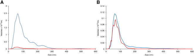
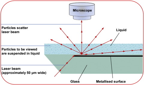
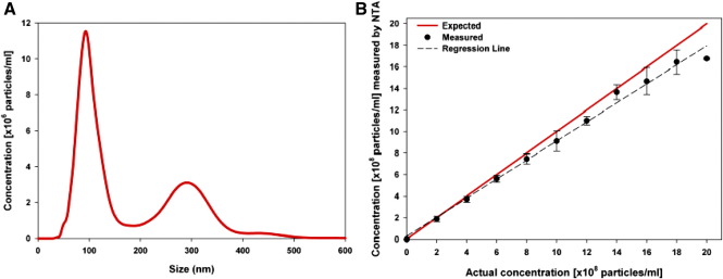
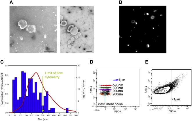
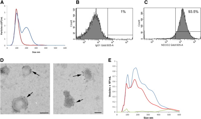
Cellular microvesicles and nanovesicles (exosomes) are involved in many disease processes and have major potential as biomarkers. However, developments in this area are constrained by limitations in the technology available for their measurement. Here we report on the use of fluorescence nanoparticle tracking analysis (NTA) to rapidly size and phenotype cellular vesicles. In this system vesicles are visualized by light scattering using a light microscope. A video is taken, and the NTA software tracks the brownian motion of individual vesicles and calculates their size and total concentration. Using human placental vesicles and plasma, we have demonstrated that NTA can measure cellular vesicles as small as ≈ 50 nm and is far more sensitive than conventional flow cytometry (lower limit ≈ 300 nm). By combining NTA with fluorescence measurement we have demonstrated that vesicles can be labeled with specific antibody-conjugated quantum dots, allowing their phenotype to be determined.
The authors of this study utilized fluorescence nanoparticle tracking analysis (NTA) to rapidly size and phenotype cellular vesicles, demonstrating that NTA is far more sensitive than conventional flow cytometry.
细胞微泡和纳米泡(外泌体)参与多种疾病过程,具有作为生物标志物的巨大潜力。然而,由于现有技术在测量这些泡体方面的局限性,该领域的发展受到了限制。在此,我们报告了使用荧光纳米颗粒跟踪分析(NTA)快速测量细胞泡体的大小和表型。在该系统中,使用显微镜通过光散射来可视化泡体。拍摄视频,然后 NTA 软件跟踪单个泡体的布朗运动并计算其大小和总浓度。我们用人胎盘泡体和血浆进行了演示,证明 NTA 可以测量小至 ≈ 50nm 的细胞泡体,比传统的流式细胞术灵敏得多(下限 ≈ 300nm)。通过将 NTA 与荧光测量相结合,我们已经证明可以用特异性抗体偶联的量子点对泡体进行标记,从而确定其表型。
本研究的作者利用荧光纳米颗粒跟踪分析(NTA)快速测量细胞泡体的大小和表型,证明 NTA 比传统的流式细胞术灵敏得多。