Mitne Somaia, Teixeira Sergio Henrique, Schwartz Michal, Belkin Michael, Farah Michel Eid, de Moraes Nilva S Bueno, da Cruz Nóia Luciana, Paes Angela Tavares, Lottenberg Cláudio Luiz, Paranhos Júnior Augusto
Department of Ophthalmology, Federal University of São Paulo.
Clin Ophthalmol. 2011;5:991-7. doi: 10.2147/OPTH.S22964. Epub 2011 Jul 15.
Evaluation of the neuroprotective effect of weekly glatiramer acetate (GA) on retinal structure and function in diabetic patients who underwent panretinal photocoagulation (PRP).
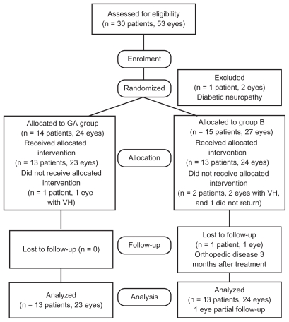
patients with severe nonproliferative or early diabetic proliferative retinopathy and no previous laser treatment were randomly divided into two groups: (1) those who received four GA treatments and (2) those who received placebo treatment. The subcutaneous injections were administered 1 week prior to laser and weekly in the subsequent three sessions of PRP in both groups. All patients underwent a full ophthalmic examination (best-corrected logMAR visual acuity, slit lamp examination, applanation tonometry, fundus biomicroscopy and indirect fundus examination); functional examination (standard automated perimetry, electroretinography and frequency-doubling technology C-20 visual field) and anatomic examination (color photography, optical coherence tomography (OCT) and Heidelberg retinal tomography). The examinations were performed before the photocoagulation and repeated 1,3,6, and 12 months after treatment (in a double-masked manner). To compare the two groups, generalized estimating equation models were performed to account for the dependence between eyes of the same patient.
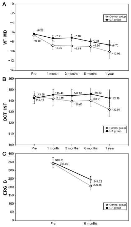
Thirteen patients (23 eyes) were included in the study group and 13 patients (24 eyes) were included in the control group. OCT showed a statistically significant difference in retinal nerve fiber layer (RNFL) thickness in the inferior peripapillary region and average thickness with thinner measurements in the control group at 1-year post-PRP. Functional analysis demonstrated a difference between groups, but it did not reach statistical significance.
The results of this study suggest that weekly GA treatment has a potential neuro-protective effect on the RNFL following photocoagulation for diabetic retinopathy.
评估每周一次醋酸格拉替雷(GA)对接受全视网膜光凝(PRP)的糖尿病患者视网膜结构和功能的神经保护作用。
患有严重非增殖性或早期糖尿病增殖性视网膜病变且既往未接受过激光治疗的患者被随机分为两组:(1)接受四次GA治疗的患者;(2)接受安慰剂治疗的患者。两组均在激光治疗前1周进行皮下注射,随后在PRP的后续三个疗程中每周注射一次。所有患者均接受了全面的眼科检查(最佳矫正对数最小分辨角视力、裂隙灯检查、压平眼压测量、眼底生物显微镜检查和间接眼底检查);功能检查(标准自动视野计、视网膜电图和倍频技术C-20视野)和解剖学检查(彩色摄影、光学相干断层扫描(OCT)和海德堡视网膜断层扫描)。检查在光凝前进行,并在治疗后1、3、6和12个月重复进行(采用双盲方式)。为比较两组,采用广义估计方程模型来考虑同一患者双眼之间的相关性。
研究组纳入13例患者(23只眼),对照组纳入13例患者(24只眼)。OCT显示,PRP后1年,对照组视乳头周围下区域视网膜神经纤维层(RNFL)厚度和平均厚度有统计学意义的差异,测量值更薄。功能分析显示两组之间存在差异,但未达到统计学意义。
本研究结果表明,每周一次的GA治疗对糖尿病性视网膜病变光凝后的RNFL具有潜在的神经保护作用。