Department of Chemistry, National University of Singapore, Science Drive 4, 117543, Singapore.
Evid Based Complement Alternat Med. 2011;2011:610625. doi: 10.1093/ecam/neq051. Epub 2011 Feb 13.
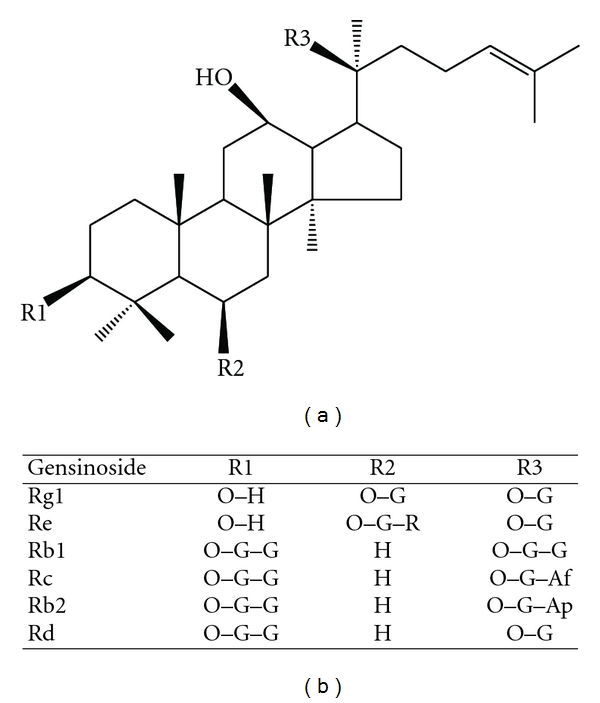
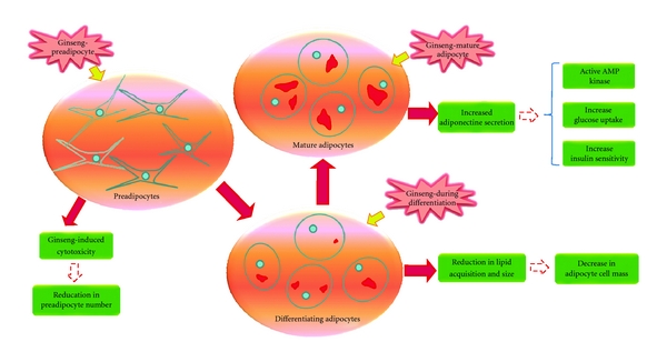
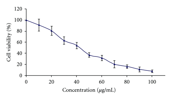
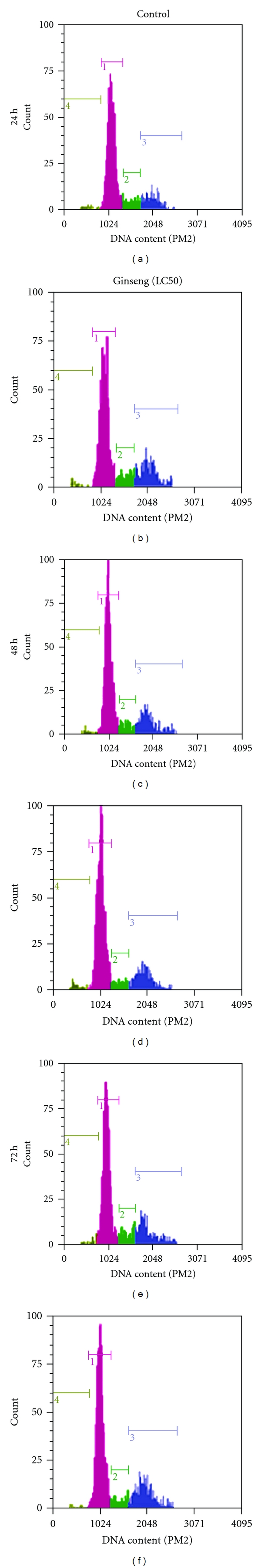
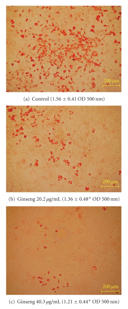
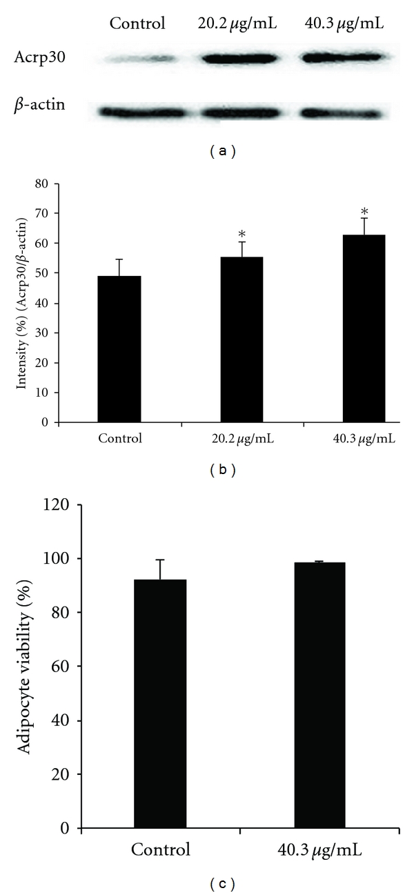
An American ginseng (Panax quinquefolius) extract (GE) that contained a quantifiable amount of ginsenosides was investigated for the potential to inhibit proliferation, affect the cell cycle, influence lipid acquisition and adiponectin expression in 3T3-L1 cells. Six fingerprint ginsenosides were quantified by high performance liquid chromatography and the respective molecular weights were confirmed by LC-ESI-MS analysis. The extract contained Rg1 (347.3 ± 99.7 μg g(-1), dry weight), Re (8280.4 ± 792.3 μg g(-1)), Rb1 (1585.8 ± 86.8 μg g(-1)), Rc (32.9 ± 8 μg g(-1)), Rb2 (62.6 ± 10.6 μg g(-1)) and Rd (90.4 ± 3.2 μg g(-1)). The GE had a dose-dependent effect on 3T3-L1 cell growth, the LC50 value was determined to be 40.3 ± 5 μg ml(-1). Cell cycle analysis showed modest changes in the cell cycle. No significant changes observed in both G1 and G2/M phases, however there was a significant decrease (P < .05) in the S phase after 24 and 48 h treatment. Apoptotic cells were modest but significantly (P < .05) increased after 48 h (3.2 ± 1.0%) compared to untreated control cells (1.5 ± 0.1%). Lipid acquisition was significantly reduced (P < .05) by 13 and 22% when treated at concentrations of 20.2 and 40.3 μg ml(-1) compared to untreated control cells. In relation to adiponectin activation, western blot analysis showed that the protein expression was significantly (P < .05) increased at concentrations tested. A quantified GE reduced the growth of 3T3-L1 cells, down-regulated the accumulation of lipid and up-regulated the expression of adiponectin in the 3T3-L1 adipocyte cell model.
一种含有可量化量的人参皂苷的西洋参(Panax quinquefolius)提取物(GE)被研究其是否具有抑制增殖、影响细胞周期、影响脂质摄取和脂联素表达的潜力。通过高效液相色谱法对 6 种指纹人参皂苷进行定量分析,并通过 LC-ESI-MS 分析分别确认各自的分子量。提取物中含有 Rg1(347.3 ± 99.7μg/g,干重)、Re(8280.4 ± 792.3μg/g)、Rb1(1585.8 ± 86.8μg/g)、Rc(32.9 ± 8μg/g)、Rb2(62.6 ± 10.6μg/g)和 Rd(90.4 ± 3.2μg/g)。GE 对 3T3-L1 细胞生长具有剂量依赖性影响,LC50 值确定为 40.3 ± 5μg/ml。细胞周期分析显示细胞周期发生了适度变化。在 G1 和 G2/M 期没有观察到显著变化,但在 24 和 48 小时处理后 S 期显著减少(P<.05)。与未处理的对照细胞(1.5 ± 0.1%)相比,凋亡细胞适度但显著增加(P<.05),48 小时后(3.2 ± 1.0%)。与未处理的对照细胞相比,当浓度分别为 20.2 和 40.3μg/ml 时,脂质摄取分别显著减少了 13%和 22%。关于脂联素激活,Western blot 分析显示,在测试的浓度下,蛋白表达显著增加(P<.05)。定量的 GE 降低了 3T3-L1 细胞的生长,下调了脂质的积累,并上调了 3T3-L1 脂肪细胞模型中脂联素的表达。