• 文献检索
  • 文档翻译
  • 深度研究
  • 学术资讯
  • Suppr Zotero 插件Zotero 插件
  • 邀请有礼
  • 套餐&价格
  • 历史记录
应用&插件
Suppr Zotero 插件Zotero 插件浏览器插件Mac 客户端Windows 客户端微信小程序
定价
高级版会员购买积分包购买API积分包
服务
文献检索文档翻译深度研究API 文档MCP 服务
关于我们
关于 Suppr公司介绍联系我们用户协议隐私条款
关注我们

Suppr 超能文献

核心技术专利:CN118964589B侵权必究
粤ICP备2023148730 号-1Suppr @ 2026

文献检索

告别复杂PubMed语法,用中文像聊天一样搜索,搜遍4000万医学文献。AI智能推荐,让科研检索更轻松。

立即免费搜索

文件翻译

保留排版,准确专业,支持PDF/Word/PPT等文件格式,支持 12+语言互译。

免费翻译文档

深度研究

AI帮你快速写综述,25分钟生成高质量综述,智能提取关键信息,辅助科研写作。

立即免费体验

基于图的医学领域分割结果的手动细化系统。

Manual refinement system for graph-based segmentation results in the medical domain.

机构信息

Department of Radiology, Harvard Medical School, Boston, MA 02115, USA.

出版信息

J Med Syst. 2012 Oct;36(5):2829-39. doi: 10.1007/s10916-011-9761-7. Epub 2011 Aug 9.

DOI:10.1007/s10916-011-9761-7
PMID:21826501
原文链接:https://pmc.ncbi.nlm.nih.gov/articles/PMC3691109/
Abstract

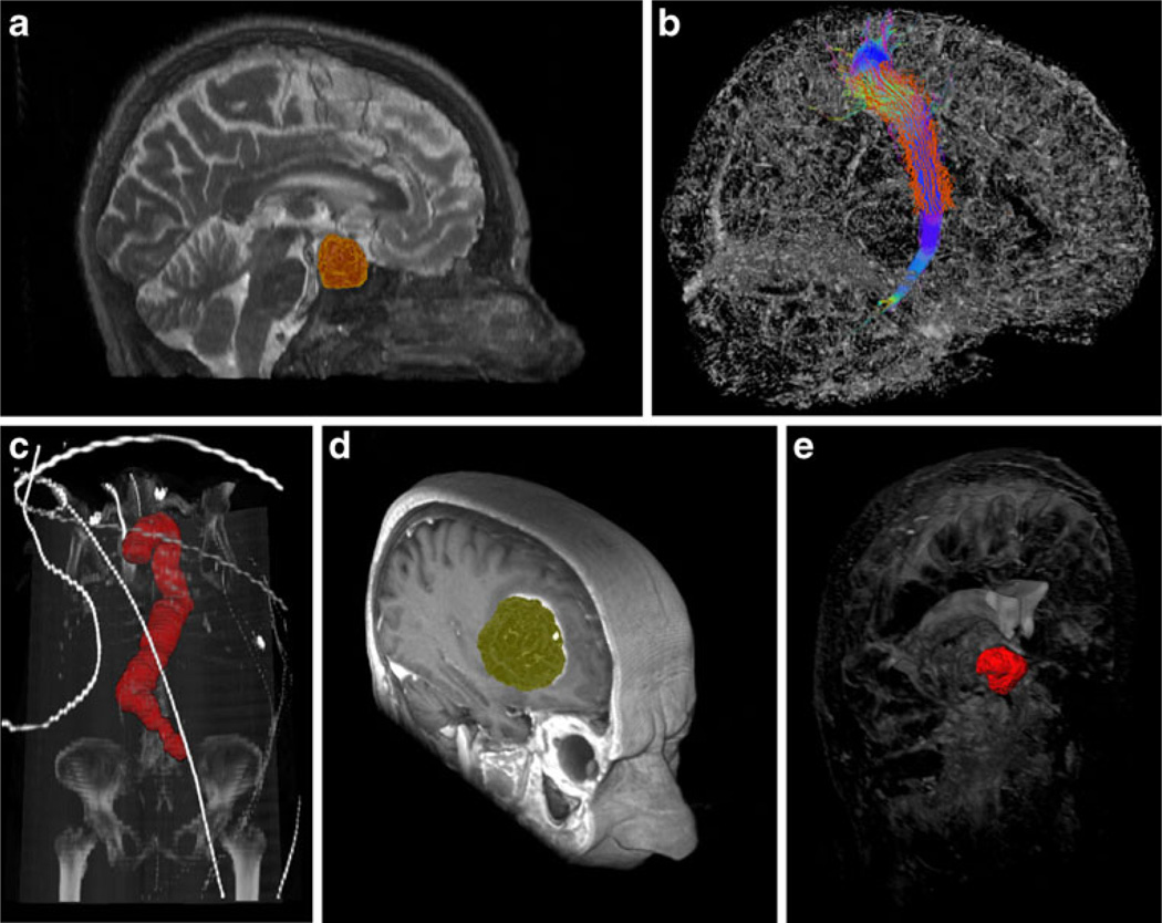
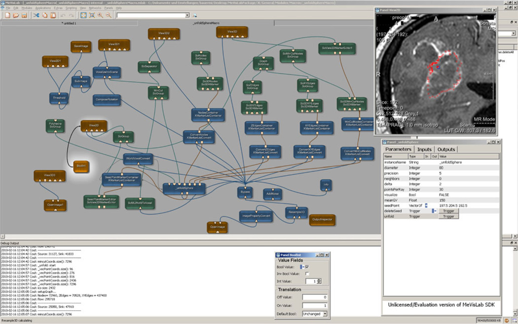
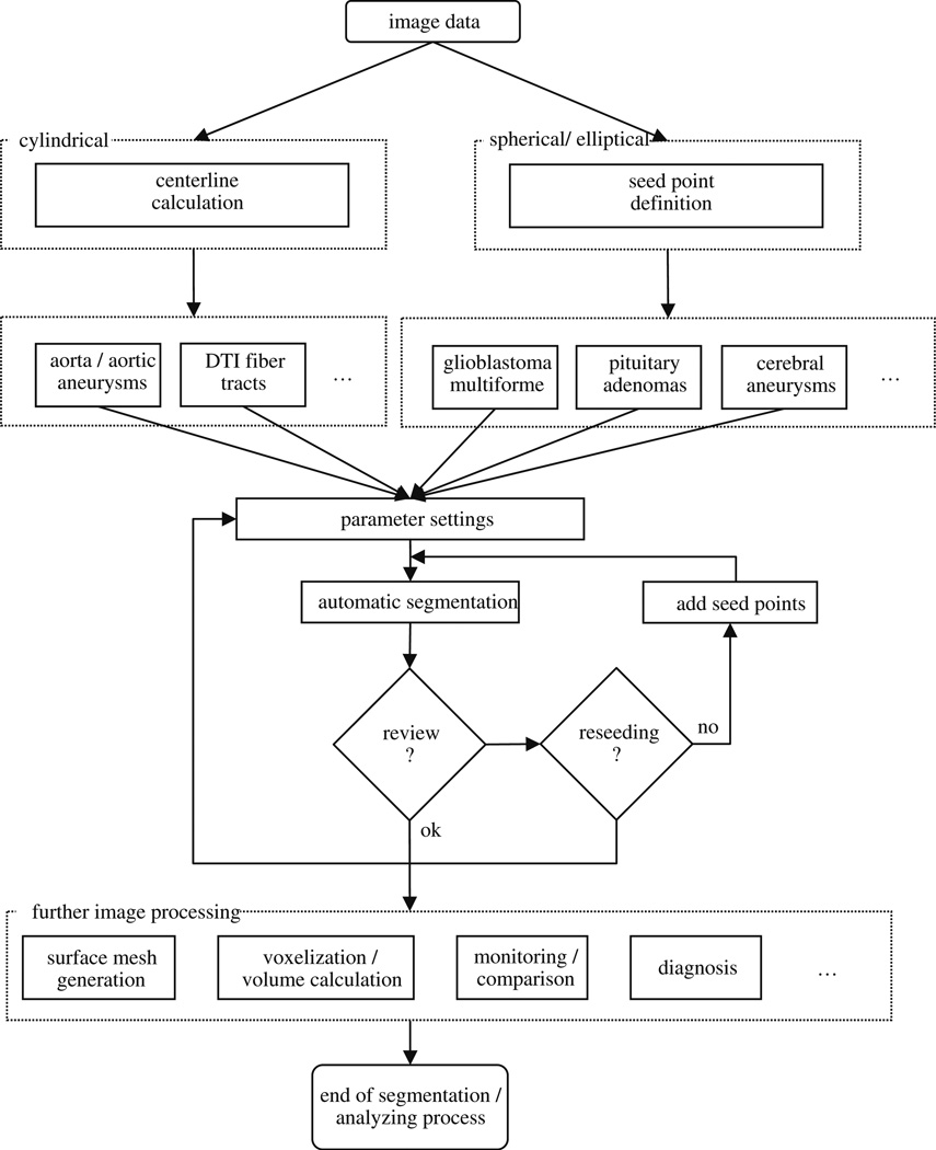
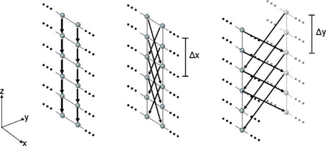
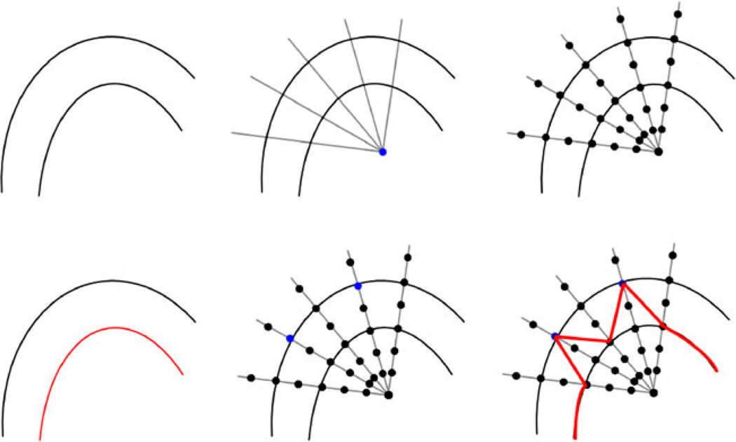
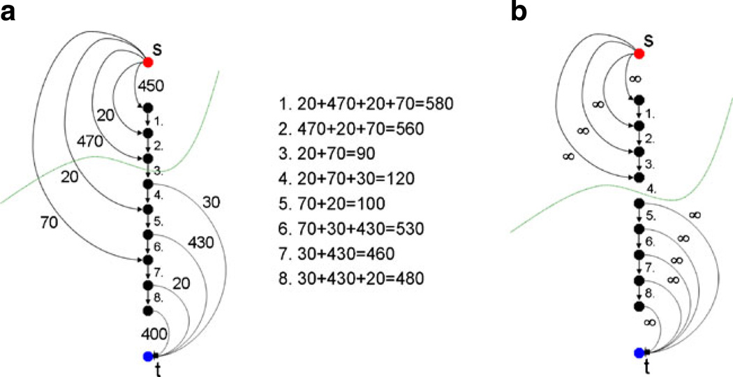
The basic principle of graph-based approaches for image segmentation is to interpret an image as a graph, where the nodes of the graph represent 2D pixels or 3D voxels of the image. The weighted edges of the graph are obtained by intensity differences in the image. Once the graph is constructed, the minimal cost closed set on the graph can be computed via a polynomial time s-t cut, dividing the graph into two parts: the object and the background. However, no segmentation method provides perfect results, so additional manual editing is required, especially in the sensitive field of medical image processing. In this study, we present a manual refinement method that takes advantage of the basic design of graph-based image segmentation algorithms. Our approach restricts a graph-cut by using additional user-defined seed points to set up fixed nodes in the graph. The advantage is that manual edits can be integrated intuitively and quickly into the segmentation result of a graph-based approach. The method can be applied to both 2D and 3D objects that have to be segmented. Experimental results for synthetic and real images are presented to demonstrate the feasibility of our approach.

摘要

基于图的图像分割方法的基本原理是将图像解释为一个图,其中图的节点表示图像的 2D 像素或 3D 体素。图的加权边由图像中的强度差异获得。一旦构建了图,就可以通过多项式时间的 s-t 割计算图上的最小代价闭集,将图分为两部分:目标和背景。然而,没有一种分割方法能提供完美的结果,因此需要进行额外的手动编辑,特别是在医学图像处理这一敏感领域。在本研究中,我们提出了一种手动细化方法,利用基于图的图像分割算法的基本设计。我们的方法通过使用附加的用户定义的种子点来限制图割,从而在图中设置固定节点。其优点是可以直观、快速地将手动编辑集成到基于图的方法的分割结果中。该方法可应用于需要分割的二维和三维物体。通过合成和真实图像的实验结果证明了我们方法的可行性。

https://cdn.ncbi.nlm.nih.gov/pmc/blobs/8fdc/3691109/b76090a402cf/nihms473377f7.jpg
https://cdn.ncbi.nlm.nih.gov/pmc/blobs/8fdc/3691109/30b3ce86f37f/nihms473377f1.jpg
https://cdn.ncbi.nlm.nih.gov/pmc/blobs/8fdc/3691109/319c6469ffb0/nihms473377f2.jpg
https://cdn.ncbi.nlm.nih.gov/pmc/blobs/8fdc/3691109/7428a9739248/nihms473377f3.jpg
https://cdn.ncbi.nlm.nih.gov/pmc/blobs/8fdc/3691109/3b8deb5463ac/nihms473377f4.jpg
https://cdn.ncbi.nlm.nih.gov/pmc/blobs/8fdc/3691109/4b77f3e11482/nihms473377f5.jpg
https://cdn.ncbi.nlm.nih.gov/pmc/blobs/8fdc/3691109/af1e3437394e/nihms473377f6.jpg
https://cdn.ncbi.nlm.nih.gov/pmc/blobs/8fdc/3691109/b76090a402cf/nihms473377f7.jpg

相似文献

1
Manual refinement system for graph-based segmentation results in the medical domain.基于图的医学领域分割结果的手动细化系统。
J Med Syst. 2012 Oct;36(5):2829-39. doi: 10.1007/s10916-011-9761-7. Epub 2011 Aug 9.
2
Interactive-cut: Real-time feedback segmentation for translational research.交互式切割:用于转化研究的实时反馈分割
Comput Med Imaging Graph. 2014 Jun;38(4):285-95. doi: 10.1016/j.compmedimag.2014.01.006. Epub 2014 Feb 11.
3
Biomedical image segmentation via constrained graph cuts and pre-segmentation.基于约束图割和预分割的生物医学图像分割
Annu Int Conf IEEE Eng Med Biol Soc. 2011;2011:5714-7. doi: 10.1109/IEMBS.2011.6091383.
4
Cube-cut: vertebral body segmentation in MRI-data through cubic-shaped divergences.立方体切割:通过立方形状散度对MRI数据中的椎体进行分割
PLoS One. 2014 Apr 4;9(4):e93389. doi: 10.1371/journal.pone.0093389. eCollection 2014.
5
A Survey of Graph Cuts/Graph Search Based Medical Image Segmentation.基于图割/图搜索的医学图像分割综述。
IEEE Rev Biomed Eng. 2018;11:112-124. doi: 10.1109/RBME.2018.2798701. Epub 2018 Jan 26.
6
Differential and relaxed image foresting transform for graph-cut segmentation of multiple 3D objects.用于多个3D物体图割分割的差分与宽松图像森林变换
Med Image Comput Comput Assist Interv. 2014;17(Pt 1):690-7. doi: 10.1007/978-3-319-10404-1_86.
7
Computer-aided lymph node segmentation in volumetric CT data.计算机辅助容积 CT 数据中的淋巴结分割。
Med Phys. 2012 Sep;39(9):5419-28. doi: 10.1118/1.4742845.
8
Pyramid graph cut: Integrating intensity and gradient information for grayscale medical image segmentation.金字塔图割:整合强度和梯度信息用于灰度医学图像分割。
Comput Biol Med. 2020 Nov;126:103997. doi: 10.1016/j.compbiomed.2020.103997. Epub 2020 Sep 19.
9
Human Part Segmentation in Depth Images with Annotated Part Positions.深度图像中带注释部位位置的人体部位分割。
Sensors (Basel). 2018 Jun 11;18(6):1900. doi: 10.3390/s18061900.
10
An energy minimization approach to the data driven editing of presegmented images/volumes.一种用于预分割图像/体数据驱动编辑的能量最小化方法。
Med Image Comput Comput Assist Interv. 2006;9(Pt 2):888-95. doi: 10.1007/11866763_109.

引用本文的文献

1
A Review of Radiomics and Artificial Intelligence and Their Application in Veterinary Diagnostic Imaging.放射组学与人工智能及其在兽医诊断成像中的应用综述
Vet Sci. 2022 Nov 8;9(11):620. doi: 10.3390/vetsci9110620.
2
Computer-aided diagnosis of cirrhosis and hepatocellular carcinoma using multi-phase abdomen CT.基于多期腹部 CT 的肝硬化和肝细胞癌计算机辅助诊断。
Int J Comput Assist Radiol Surg. 2019 Aug;14(8):1341-1352. doi: 10.1007/s11548-019-01991-5. Epub 2019 May 6.
3
Computed tomography data collection of the complete human mandible and valid clinical ground truth models.

本文引用的文献

1
A medical software system for volumetric analysis of cerebral pathologies in magnetic resonance imaging (MRI) data.用于磁共振成像 (MRI) 数据中脑病理学容积分析的医学软件系统。
J Med Syst. 2012 Aug;36(4):2097-109. doi: 10.1007/s10916-011-9673-6. Epub 2011 Mar 8.
2
Aortic volume as an indicator of disease progression in patients with untreated infrarenal abdominal aneurysm.主动脉体积作为未经治疗的肾下型腹主动脉瘤患者疾病进展的指标。
Eur J Radiol. 2012 Feb;81(2):e87-93. doi: 10.1016/j.ejrad.2011.01.077. Epub 2011 Feb 12.
3
Optimal surface segmentation in volumetric images--a graph-theoretic approach.
全下颌骨的计算机断层扫描数据采集和有效的临床地面真实模型。
Sci Data. 2019 Jan 29;6:190003. doi: 10.1038/sdata.2019.3.
4
Clinical evaluation of semi-automatic open-source algorithmic software segmentation of the mandibular bone: Practical feasibility and assessment of a new course of action.临床评估下颌骨半自动开源算法软件分割:实用可行性及新行动方案评估。
PLoS One. 2018 May 10;13(5):e0196378. doi: 10.1371/journal.pone.0196378. eCollection 2018.
5
A Segmentation Editing Framework Based on Shape Change Statistics.一种基于形状变化统计的分割编辑框架。
Proc SPIE Int Soc Opt Eng. 2017;10133. doi: 10.1117/12.2250023. Epub 2017 Feb 24.
6
Iterative-cuts: longitudinal and scale-invariant segmentation via user-defined templates for rectosigmoid colon in gynecological brachytherapy.迭代切割:通过用户定义模板对妇科近距离放射治疗中的直肠乙状结肠进行纵向和尺度不变分割。
J Med Imaging (Bellingham). 2016 Apr;3(2):024004. doi: 10.1117/1.JMI.3.2.024004. Epub 2016 Jun 20.
7
A Novel Active Contour Model for MRI Brain Segmentation used in Radiotherapy Treatment Planning.一种用于放射治疗计划的磁共振成像脑部分割的新型活动轮廓模型。
Electron Physician. 2016 May 25;8(5):2443-51. doi: 10.19082/2443. eCollection 2016 May.
8
Interactive Volumetry Of Liver Ablation Zones.肝脏消融区的交互式容积测量
Sci Rep. 2015 Oct 20;5:15373. doi: 10.1038/srep15373.
9
Brain MR image segmentation with spatial constrained K-mean algorithm and dual-tree complex wavelet transform.基于空间约束K均值算法和双树复小波变换的脑磁共振图像分割
J Med Syst. 2014 Sep;38(9):93. doi: 10.1007/s10916-014-0093-2. Epub 2014 Jul 4.
10
Refinement-cut: user-guided segmentation algorithm for translational science.精细化切割:用于转化医学的用户引导分割算法
Sci Rep. 2014 Jun 4;4:5164. doi: 10.1038/srep05164.
体积图像中的最优表面分割——一种基于图论的方法。
IEEE Trans Pattern Anal Mach Intell. 2006 Jan;28(1):119-34. doi: 10.1109/TPAMI.2006.19.
4
An experimental comparison of min-cut/max-flow algorithms for energy minimization in vision.用于视觉能量最小化的最小割/最大流算法的实验比较。
IEEE Trans Pattern Anal Mach Intell. 2004 Sep;26(9):1124-37. doi: 10.1109/TPAMI.2004.60.
5
Statistical validation of image segmentation quality based on a spatial overlap index.基于空间重叠指数的图像分割质量的统计验证。
Acad Radiol. 2004 Feb;11(2):178-89. doi: 10.1016/s1076-6332(03)00671-8.
6
Current methods in medical image segmentation.医学图像分割的当前方法。
Annu Rev Biomed Eng. 2000;2:315-37. doi: 10.1146/annurev.bioeng.2.1.315.