Kapanci Y, Burgan S, Pietra G G, Conne B, Gabbiani G
Department of Pathology, University of Geneva, Switzerland.
Am J Pathol. 1990 Apr;136(4):881-9.
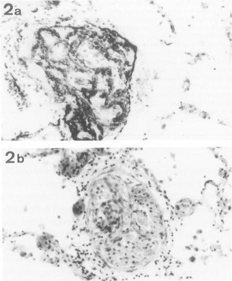
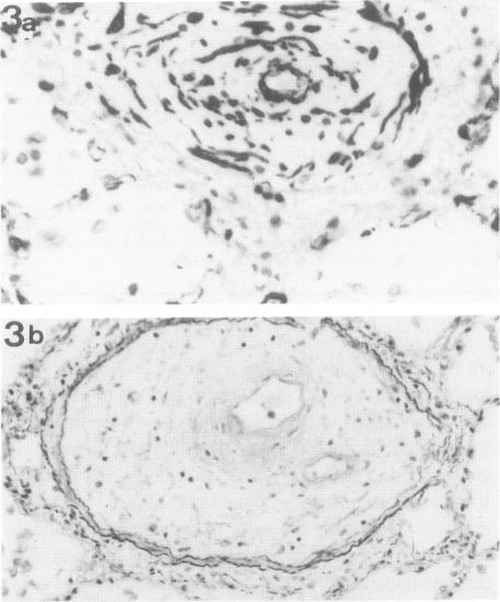
Lungs of 37 patients with pulmonary hypertension (PHT), 5 normal human lungs, and 30 normal rat lungs, were studied using immunohistochemical stainings for actin, alpha-smooth muscle (alpha-SM) actin and desmin. The type of PHT was determined on clinicopathologic grounds (in 17 cases by catheterism); 20 patients had precapillary and 17 postcapillary PHT. In normal lungs, myofibroblasts, ie, contractile interstitial cells (CIC), distributed in alveolar septa, were not stained by alpha-SM actin antibodies. Only around the venules, were cells labeled by this antibody present. Furthermore, there were bundles of alpha-SM actin-positive cells around the openings of air sacculi into the alveolar ducts. In precapillary PHT, the distribution and immunostaining properties of interstitial cells remained unchanged; alpha-SM actin-positive cells were observed in thickened arterial intima and in plexiform lesions. In postcapillary PHT secondary to heart failure, to mitral stenosis, or in veno-occlusive disease, many interstitial cells in the alveolar septa were decorated by alpha-SM actin antibodies but not with desmin. The authors propose that, in postcapillary PHT, mechanical stretch due to capillary congestion may be responsible for the generation of cells that express an actin isoform associated with smooth muscle.
对37例肺动脉高压(PHT)患者的肺、5个正常人类肺组织以及30个正常大鼠肺组织进行了研究,采用肌动蛋白、α-平滑肌(α-SM)肌动蛋白和结蛋白的免疫组织化学染色。PHT的类型根据临床病理依据确定(17例通过心导管检查);20例患者为毛细血管前性PHT,17例为毛细血管后性PHT。在正常肺组织中,肌成纤维细胞,即收缩性间质细胞(CIC),分布于肺泡隔,未被α-SM肌动蛋白抗体染色。仅在小静脉周围有被该抗体标记的细胞。此外,在肺泡囊开口进入肺泡管处周围有α-SM肌动蛋白阳性细胞束。在毛细血管前性PHT中,间质细胞的分布和免疫染色特性保持不变;在增厚的动脉内膜和丛状病变中观察到α-SM肌动蛋白阳性细胞。在继发于心力衰竭、二尖瓣狭窄或静脉闭塞性疾病的毛细血管后性PHT中,肺泡隔中的许多间质细胞被α-SM肌动蛋白抗体标记,但未被结蛋白标记。作者提出,在毛细血管后性PHT中,毛细血管充血引起的机械性牵张可能是导致表达与平滑肌相关的肌动蛋白异构体的细胞产生的原因。