• 文献检索
  • 文档翻译
  • 深度研究
  • 学术资讯
  • Suppr Zotero 插件Zotero 插件
  • 邀请有礼
  • 套餐&价格
  • 历史记录
应用&插件
Suppr Zotero 插件Zotero 插件浏览器插件Mac 客户端Windows 客户端微信小程序
定价
高级版会员购买积分包购买API积分包
服务
文献检索文档翻译深度研究API 文档MCP 服务
关于我们
关于 Suppr公司介绍联系我们用户协议隐私条款
关注我们

Suppr 超能文献

核心技术专利:CN118964589B侵权必究
粤ICP备2023148730 号-1Suppr @ 2026

文献检索

告别复杂PubMed语法,用中文像聊天一样搜索,搜遍4000万医学文献。AI智能推荐,让科研检索更轻松。

立即免费搜索

文件翻译

保留排版,准确专业,支持PDF/Word/PPT等文件格式,支持 12+语言互译。

免费翻译文档

深度研究

AI帮你快速写综述,25分钟生成高质量综述,智能提取关键信息,辅助科研写作。

立即免费体验

相似文献

1
Computerized three-class classification of MRI-based prognostic markers for breast cancer.基于 MRI 的乳腺癌预后标志物的计算机化三分类。
Phys Med Biol. 2011 Sep 21;56(18):5995-6008. doi: 10.1088/0031-9155/56/18/014. Epub 2011 Aug 22.
2
Potential of computer-aided diagnosis of high spectral and spatial resolution (HiSS) MRI in the classification of breast lesions.高光谱和空间分辨率(HiSS)MRI 计算机辅助诊断在乳腺病变分类中的应用潜力。
J Magn Reson Imaging. 2014 Jan;39(1):59-67. doi: 10.1002/jmri.24145. Epub 2013 Sep 10.
3
Multimodality computer-aided breast cancer diagnosis with FFDM and DCE-MRI.基于 FFDM 和 DCE-MRI 的多模态计算机辅助乳腺癌诊断。
Acad Radiol. 2010 Sep;17(9):1158-67. doi: 10.1016/j.acra.2010.04.015.
4
A computer-aided diagnosis system for breast DCE-MRI at high spatiotemporal resolution.一种用于高时空分辨率乳腺动态对比增强磁共振成像的计算机辅助诊断系统。
Med Phys. 2016 Jan;43(1):84. doi: 10.1118/1.4937787.
5
Cancerous breast lesions on dynamic contrast-enhanced MR images: computerized characterization for image-based prognostic markers.动态对比增强磁共振图像上的癌性乳腺病变:基于图像的预后标志物的计算机特征化。
Radiology. 2010 Mar;254(3):680-90. doi: 10.1148/radiol.09090838. Epub 2010 Feb 1.
6
Multimodality computerized diagnosis of breast lesions using mammography and sonography.使用乳腺X线摄影和超声检查对乳腺病变进行多模态计算机诊断。
Acad Radiol. 2005 Aug;12(8):970-9. doi: 10.1016/j.acra.2005.04.014.
7
Role of sureness in evaluating AI/CADx: Lesion-based repeatability of machine learning classification performance on breast MRI.Surety 在评估 AI/CADx 中的作用:基于病灶的机器学习分类性能在乳腺 MRI 上的重复性。
Med Phys. 2024 Mar;51(3):1812-1821. doi: 10.1002/mp.16673. Epub 2023 Aug 21.
8
Computerized assessment of breast lesion malignancy using DCE-MRI robustness study on two independent clinical datasets from two manufacturers.使用来自两个制造商的两个独立临床数据集的 DCE-MRI 稳健性研究对乳腺病变恶性程度进行计算机评估。
Acad Radiol. 2010 Jul;17(7):822-9. doi: 10.1016/j.acra.2010.03.007.
9
Integration of dynamic contrast-enhanced magnetic resonance imaging and T2-weighted imaging radiomic features by a canonical correlation analysis-based feature fusion method to predict histological grade in ductal breast carcinoma.基于典型相关分析的特征融合方法整合动态对比增强磁共振成像和 T2 加权成像放射组学特征,以预测导管乳腺癌的组织学分级。
Phys Med Biol. 2019 Oct 23;64(21):215001. doi: 10.1088/1361-6560/ab3fd3.
10
Combined use of T2-weighted MRI and T1-weighted dynamic contrast-enhanced MRI in the automated analysis of breast lesions.T2 加权 MRI 与 T1 加权动态对比增强 MRI 在自动分析乳腺病变中的联合应用。
Magn Reson Med. 2011 Aug;66(2):555-64. doi: 10.1002/mrm.22800. Epub 2011 Apr 26.

引用本文的文献

1
Sureness of classification of breast cancers as pure ductal carcinoma or with invasive components on dynamic contrast-enhanced magnetic resonance imaging: application of likelihood assurance metrics for computer-aided diagnosis.动态对比增强磁共振成像中乳腺癌分类为纯导管癌或伴有浸润成分的确定性:用于计算机辅助诊断的似然性保证指标的应用
J Med Imaging (Bellingham). 2025 Nov;12(Suppl 2):S22012. doi: 10.1117/1.JMI.12.S2.S22012. Epub 2025 Jun 18.
2
Characterization of Breast Tumors from MR Images Using Radiomics and Machine Learning Approaches.使用影像组学和机器学习方法对乳腺肿瘤进行特征分析。
J Pers Med. 2023 Jun 28;13(7):1062. doi: 10.3390/jpm13071062.
3
Predicting Malignancy of Breast Imaging Findings Using Quantitative Analysis of Contrast-Enhanced Mammography (CEM).使用对比增强乳腺X线摄影(CEM)的定量分析预测乳腺影像结果的恶性程度
Diagnostics (Basel). 2023 Mar 16;13(6):1129. doi: 10.3390/diagnostics13061129.
4
Invasive ductal breast cancer molecular subtype prediction by MRI radiomic and clinical features based on machine learning.基于机器学习,通过MRI影像组学和临床特征预测浸润性导管乳腺癌分子亚型
Front Oncol. 2022 Sep 12;12:964605. doi: 10.3389/fonc.2022.964605. eCollection 2022.
5
Radiomics for Tumor Characterization in Breast Cancer Patients: A Feasibility Study Comparing Contrast-Enhanced Mammography and Magnetic Resonance Imaging.用于乳腺癌患者肿瘤特征描述的放射组学:一项对比增强乳腺X线摄影和磁共振成像的可行性研究
Diagnostics (Basel). 2020 Jul 18;10(7):492. doi: 10.3390/diagnostics10070492.
6
The Impact of Normalization Approaches to Automatically Detect Radiogenomic Phenotypes Characterizing Breast Cancer Receptors Status.标准化方法对自动检测表征乳腺癌受体状态的放射基因组表型的影响。
Cancers (Basel). 2020 Feb 24;12(2):518. doi: 10.3390/cancers12020518.
7
Radiogenomics of breast cancer using dynamic contrast enhanced MRI and gene expression profiling.基于动态对比增强 MRI 和基因表达谱的乳腺癌放射组学研究。
Cancer Imaging. 2019 Jul 15;19(1):48. doi: 10.1186/s40644-019-0233-5.
8
Predicting Breast Cancer Molecular Subtype with MRI Dataset Utilizing Convolutional Neural Network Algorithm.利用卷积神经网络算法对 MRI 数据集进行乳腺癌分子亚型预测。
J Digit Imaging. 2019 Apr;32(2):276-282. doi: 10.1007/s10278-019-00179-2.
9
Relationships Between Human-Extracted MRI Tumor Phenotypes of Breast Cancer and Clinical Prognostic Indicators Including Receptor Status and Molecular Subtype.人类提取的乳腺癌MRI肿瘤表型与包括受体状态和分子亚型在内的临床预后指标之间的关系。
Curr Probl Diagn Radiol. 2019 Sep-Oct;48(5):467-472. doi: 10.1067/j.cpradiol.2018.08.003. Epub 2018 Aug 23.
10
Breast MRI radiomics: comparison of computer- and human-extracted imaging phenotypes.乳腺MRI影像组学:计算机提取与人工提取影像表型的比较
Eur Radiol Exp. 2017;1(1):22. doi: 10.1186/s41747-017-0025-2. Epub 2017 Nov 21.

本文引用的文献

1
Evaluation of clinical breast MR imaging performed with prototype computer-aided diagnosis breast MR imaging workstation: reader study.应用原型计算机辅助诊断乳腺磁共振成像工作站行乳腺磁共振成像的临床评估:读者研究。
Radiology. 2011 Mar;258(3):696-704. doi: 10.1148/radiol.10100409. Epub 2011 Jan 6.
2
Cancer statistics, 2010.癌症统计数据,2010 年。
CA Cancer J Clin. 2010 Sep-Oct;60(5):277-300. doi: 10.3322/caac.20073. Epub 2010 Jul 7.
3
Cancerous breast lesions on dynamic contrast-enhanced MR images: computerized characterization for image-based prognostic markers.动态对比增强磁共振图像上的癌性乳腺病变:基于图像的预后标志物的计算机特征化。
Radiology. 2010 Mar;254(3):680-90. doi: 10.1148/radiol.09090838. Epub 2010 Feb 1.
4
Predicting survival and early clinical response to primary chemotherapy for patients with locally advanced breast cancer using DCE-MRI.使用动态对比增强磁共振成像(DCE-MRI)预测局部晚期乳腺癌患者对原发性化疗的生存情况和早期临床反应。
J Magn Reson Imaging. 2009 Jun;29(6):1300-7. doi: 10.1002/jmri.21778.
5
Anniversary paper: History and status of CAD and quantitative image analysis: the role of Medical Physics and AAPM.周年纪念论文:冠心病及定量图像分析的历史与现状:医学物理与美国医学物理学家协会的作用
Med Phys. 2008 Dec;35(12):5799-820. doi: 10.1118/1.3013555.
6
The impact of preoperative MRI on breast-conserving surgery of invasive cancer: a comparative cohort study.术前磁共振成像对浸润性癌保乳手术的影响:一项比较队列研究。
Breast Cancer Res Treat. 2009 Jul;116(1):161-9. doi: 10.1007/s10549-008-0182-3. Epub 2008 Sep 21.
7
Breast MRI for cancer detection and characterization: a review of evidence-based clinical applications.用于癌症检测与特征分析的乳腺磁共振成像:基于证据的临床应用综述
Acad Radiol. 2008 Apr;15(4):408-16. doi: 10.1016/j.acra.2007.11.006.
8
Volumetric texture analysis of breast lesions on contrast-enhanced magnetic resonance images.对比增强磁共振图像上乳腺病变的容积纹理分析
Magn Reson Med. 2007 Sep;58(3):562-71. doi: 10.1002/mrm.21347.
9
An overview of prognostic factors for long-term survivors of breast cancer.乳腺癌长期幸存者的预后因素概述。
Breast Cancer Res Treat. 2008 Feb;107(3):309-30. doi: 10.1007/s10549-007-9556-1. Epub 2007 Mar 22.
10
Computer assistance for MR based diagnosis of breast cancer: present and future challenges.基于磁共振成像的乳腺癌诊断的计算机辅助:当前与未来的挑战
Comput Med Imaging Graph. 2007 Jun-Jul;31(4-5):236-47. doi: 10.1016/j.compmedimag.2007.02.007. Epub 2007 Mar 21.

基于 MRI 的乳腺癌预后标志物的计算机化三分类。

Computerized three-class classification of MRI-based prognostic markers for breast cancer.

机构信息

Department of Radiology, The University of Chicago, Chicago, IL 60637, USA.

出版信息

Phys Med Biol. 2011 Sep 21;56(18):5995-6008. doi: 10.1088/0031-9155/56/18/014. Epub 2011 Aug 22.

DOI:10.1088/0031-9155/56/18/014
PMID:21860079
原文链接:https://pmc.ncbi.nlm.nih.gov/articles/PMC4134441/
Abstract

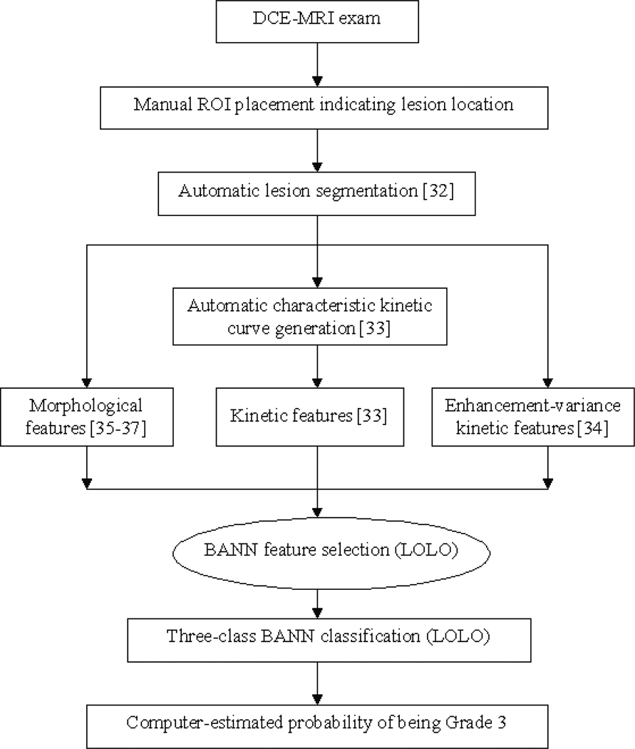
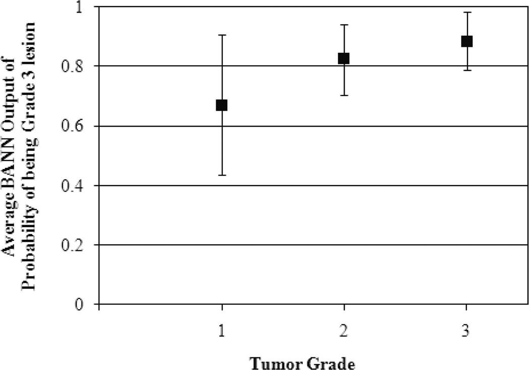
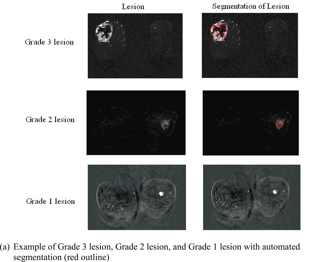
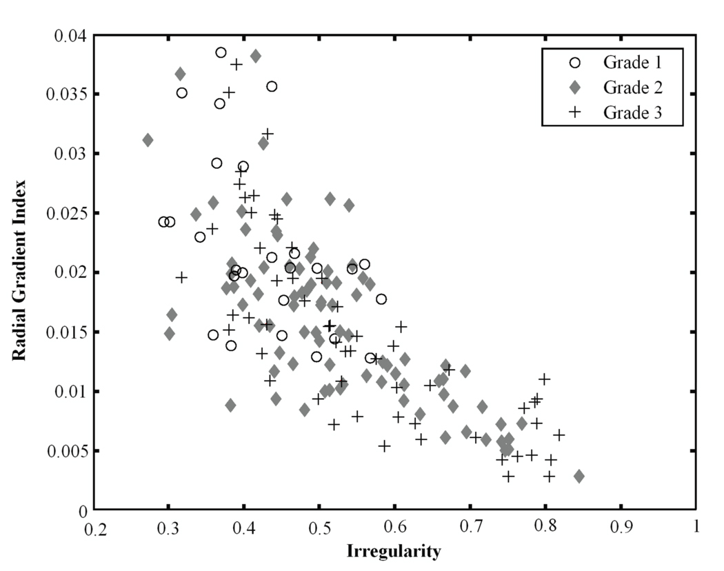
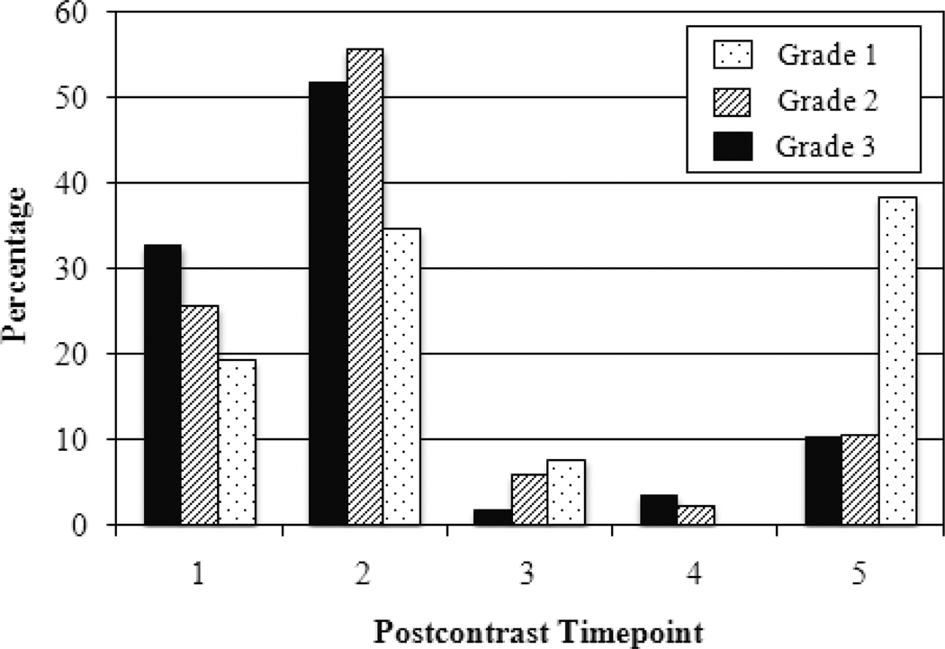
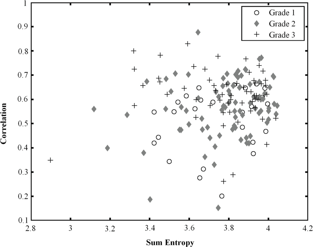
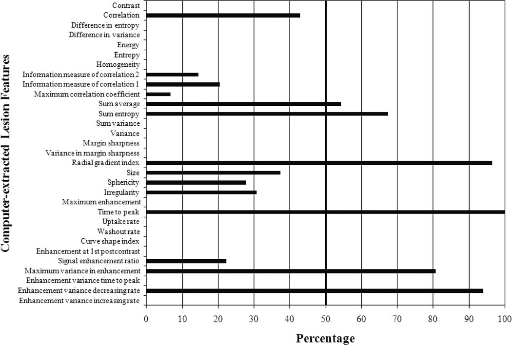
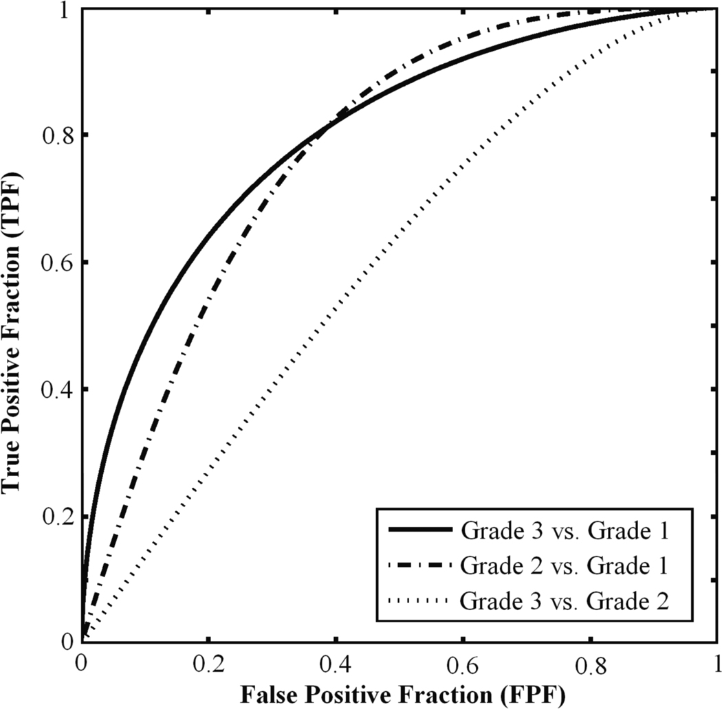
The purpose of this study is to investigate whether computerized analysis using three-class Bayesian artificial neural network (BANN) feature selection and classification can characterize tumor grades (grade 1, grade 2 and grade 3) of breast lesions for prognostic classification on DCE-MRI. A database of 26 IDC grade 1 lesions, 86 IDC grade 2 lesions and 58 IDC grade 3 lesions was collected. The computer automatically segmented the lesions, and kinetic and morphological lesion features were automatically extracted. The discrimination tasks-grade 1 versus grade 3, grade 2 versus grade 3, and grade 1 versus grade 2 lesions-were investigated. Step-wise feature selection was conducted by three-class BANNs. Classification was performed with three-class BANNs using leave-one-lesion-out cross-validation to yield computer-estimated probabilities of being grade 3 lesion, grade 2 lesion and grade 1 lesion. Two-class ROC analysis was used to evaluate the performances. We achieved AUC values of 0.80 ± 0.05, 0.78 ± 0.05 and 0.62 ± 0.05 for grade 1 versus grade 3, grade 1 versus grade 2, and grade 2 versus grade 3, respectively. This study shows the potential for (1) applying three-class BANN feature selection and classification to CADx and (2) expanding the role of DCE-MRI CADx from diagnostic to prognostic classification in distinguishing tumor grades.

摘要

本研究旨在探讨基于三分类贝叶斯人工神经网络(BANN)特征选择和分类的计算机分析是否可用于对 DCE-MRI 中的乳腺病变进行肿瘤分级(1 级、2 级和 3 级)的预后分类。收集了 26 个 IDC 1 级病变、86 个 IDC 2 级病变和 58 个 IDC 3 级病变的数据库。计算机自动对病变进行分割,自动提取动力学和形态学病变特征。研究了鉴别任务——1 级与 3 级、2 级与 3 级和 1 级与 2 级病变。通过三分类 BANN 进行逐步特征选择。使用三分类 BANN 进行分类,采用留一病变交叉验证,得出计算机估计的病变为 3 级、2 级和 1 级的概率。采用二分类 ROC 分析评估性能。对于 1 级与 3 级、1 级与 2 级和 2 级与 3 级的病变,AUC 值分别为 0.80±0.05、0.78±0.05 和 0.62±0.05。本研究表明,(1)应用三分类 BANN 特征选择和分类用于 CADx 具有潜力,(2)DCE-MRI CADx 从诊断扩展到肿瘤分级的预后分类的作用。

https://cdn.ncbi.nlm.nih.gov/pmc/blobs/9549/4134441/abb5628dc45a/nihms321898f8.jpg
https://cdn.ncbi.nlm.nih.gov/pmc/blobs/9549/4134441/9d79501d27a2/nihms321898f1.jpg
https://cdn.ncbi.nlm.nih.gov/pmc/blobs/9549/4134441/e492e9d1fa38/nihms321898f2a.jpg
https://cdn.ncbi.nlm.nih.gov/pmc/blobs/9549/4134441/6cf6415336a1/nihms321898f3.jpg
https://cdn.ncbi.nlm.nih.gov/pmc/blobs/9549/4134441/e0592dbdfed9/nihms321898f4.jpg
https://cdn.ncbi.nlm.nih.gov/pmc/blobs/9549/4134441/30617ad1f8da/nihms321898f5.jpg
https://cdn.ncbi.nlm.nih.gov/pmc/blobs/9549/4134441/6ffc4d833343/nihms321898f6.jpg
https://cdn.ncbi.nlm.nih.gov/pmc/blobs/9549/4134441/b28af302868e/nihms321898f7.jpg
https://cdn.ncbi.nlm.nih.gov/pmc/blobs/9549/4134441/abb5628dc45a/nihms321898f8.jpg